


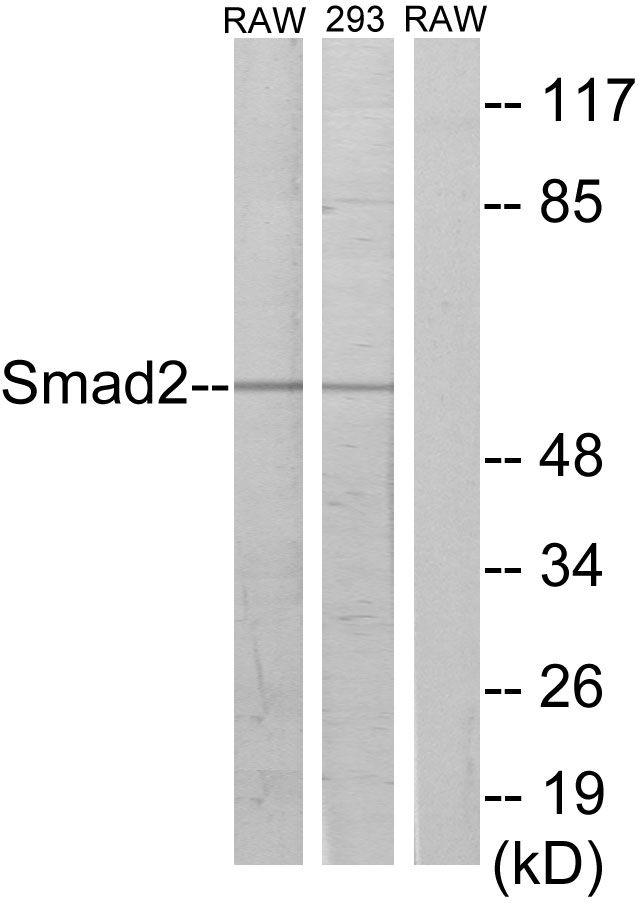
Figure 1. Western blot analysis of SMAD2 using anti-SMAD2 antibody (A00090-1). Electrophoresis was performed on a 5-20% SDS-PAGE gel at 70V (Stacking gel) / 90V (Resolving gel) for 2-3 hours. The sample well of each lane was loaded with 50ug of sample under reducing conditions. Lane 1: human placenta tissue lysates Lane 2: rat liver tissue lysates Lane 3: mouse testicular tissue lysates Lane 4: mouse heart tissue lysates Lane 5: mouse lung tissue lysates Lane 6: mouse lung tissue lysates After Electrophoresis, proteins were transferred to a Nitrocellulose membrane at 150mA for 50-90 minutes. Blocked the membrane with 5% Non-fat Milk/ TBS for 1.5 hour at RT. The membrane was incubated with rabbit anti-SMAD2 antigen affinity purified polyclonal antibody (Catalog # A00090-1) at 0.5 microg/mL overnight at 4°C, then washed with TBS-0.1%Tween 3 times with 5 minutes each and probed with a goat anti-rabbit IgG-HRP secondary antibody at a dilution of 1:10000 for 1.5 hour at RT. The signal is developed using an Enhanced Chemiluminescent detection (ECL) kit (Catalog # EK1002) with Tanon 5200 system. A specific band was detected for SMAD2 at approximately 60KD. The expected band size for SMAD2 is at 52KD.
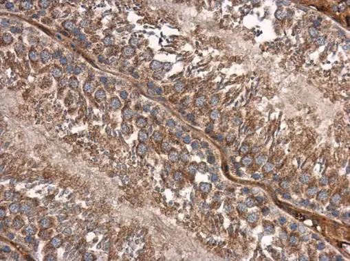
. SMAD2 was detected in paraffin-embedded section of human colon cancer tissues. Heat mediated antigen retrieval was performed in citrate buffer (pH6, epitope retrieval solution) for 20 mins. The tissue section was blocked with 10% goat serum. The tissue section was then incubated with 1microg/ml rabbit anti-SMAD2 Antibody (A00090-1) overnight at 4°C. Biotinylated goat anti-rabbit IgG was used as secondary antibody and incubated for 30 minutes at 37°C. The tissue section was developed using Strepavidin-Biotin-Complex (SABC)(Catalog # SA1022) with DAB as the chromogen.")
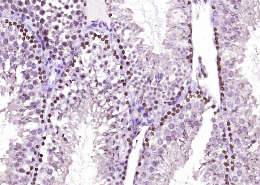
. SMAD2 was detected in paraffin-embedded section of human placenta tissues. Heat mediated antigen retrieval was performed in citrate buffer (pH6, epitope retrieval solution) for 20 mins. The tissue section was blocked with 10% goat serum. The tissue section was then incubated with 1microg/ml rabbit anti-SMAD2 Antibody (A00090-1) overnight at 4°C. Biotinylated goat anti-rabbit IgG was used as secondary antibody and incubated for 30 minutes at 37°C. The tissue section was developed using Strepavidin-Biotin-Complex (SABC)(Catalog # SA1022) with DAB as the chromogen.")
. SMAD2 was detected in paraffin-embedded section of mouse brain tissues. Heat mediated antigen retrieval was performed in citrate buffer (pH6, epitope retrieval solution) for 20 mins. The tissue section was blocked with 10% goat serum. The tissue section was then incubated with 1microg/ml rabbit anti-SMAD2 Antibody (A00090-1) overnight at 4°C. Biotinylated goat anti-rabbit IgG was used as secondary antibody and incubated for 30 minutes at 37°C. The tissue section was developed using Strepavidin-Biotin-Complex (SABC)(Catalog # SA1022) with DAB as the chromogen.")
. SMAD2 was detected in paraffin-embedded section of rat brain tissues. Heat mediated antigen retrieval was performed in citrate buffer (pH6, epitope retrieval solution) for 20 mins. The tissue section was blocked with 10% goat serum. The tissue section was then incubated with 1microg/ml rabbit anti-SMAD2 Antibody (A00090-1) overnight at 4°C. Biotinylated goat anti-rabbit IgG was used as secondary antibody and incubated for 30 minutes at 37°C. The tissue section was developed using Strepavidin-Biotin-Complex (SABC)(Catalog # SA1022) with DAB as the chromogen.")
. SMAD2 was detected in immunocytochemical section of HELA cells. Enzyme antigen retrieval was performed using IHC enzyme antigen retrieval reagent (AR0022) for 15 mins. The cells were blocked with 10% goat serum. And then incubated with 5microg/mL rabbit anti-SMAD2 Antibody (A00090-1) overnight at 4°C. DyLight®594 Conjugated Goat Anti-Rabbit IgG (BA1142) was used as secondary antibody at 1:100 dilution and incubated for 30 minutes at 37°C. The section was counterstained with DAPI. Visualize using a fluorescence microscope and filter sets appropriate for the label used.")
. Overlay histogram showing K562 cells stained with A00090-1 (Blue line).To facilitate intracellular staining, cells were fixed with 4% paraformaldehyde and permeabilized with permeabilization buffer. The cells were blocked with 10% normal goat serum. And then incubated with rabbit anti-SMAD2 Antibody (A00090-1, 1microg/1x106 cells) for 30 min at 20°C. DyLight®488 conjugated goat anti-rabbit IgG (BA1127, 5-10microg/1x106 cells) was used as secondary antibody for 30 minutes at 20°C. Isotype control antibody (Green line) was rabbit IgG (1microg/1x106) used under the same conditions. Unlabelled sample without incubation with primary antibody and secondary antibody (Red line) was used as a blank control.")
Figure 1. Western blot analysis of SMAD2 using anti-SMAD2 antibody (A00090-1). Electrophoresis was performed on a 5-20% SDS-PAGE gel at 70V (Stacking gel) / 90V (Resolving gel) for 2-3 hours. The sample well of each lane was loaded with 50ug of sample under reducing conditions. Lane 1: human placenta tissue lysates Lane 2: rat liver tissue lysates Lane 3: mouse testicular tissue lysates Lane 4: mouse heart tissue lysates Lane 5: mouse lung tissue lysates Lane 6: mouse lung tissue lysates After Electrophoresis, proteins were transferred to a Nitrocellulose membrane at 150mA for 50-90 minutes. Blocked the membrane with 5% Non-fat Milk/ TBS for 1.5 hour at RT. The membrane was incubated with rabbit anti-SMAD2 antigen affinity purified polyclonal antibody (Catalog # A00090-1) at 0.5 microg/mL overnight at 4°C, then washed with TBS-0.1%Tween 3 times with 5 minutes each and probed with a goat anti-rabbit IgG-HRP secondary antibody at a dilution of 1:10000 for 1.5 hour at RT. The signal is developed using an Enhanced Chemiluminescent detection (ECL) kit (Catalog # EK1002) with Tanon 5200 system. A specific band was detected for SMAD2 at approximately 60KD. The expected band size for SMAD2 is at 52KD.
Anti-SMAD2 Antibody Picoband(r)
A00090-1-CARRIER-FREE
ApplicationsFlow Cytometry, ImmunoFluorescence, Western Blot, ELISA, ImmunoCytoChemistry, ImmunoHistoChemistry
Product group Antibodies
ReactivityHuman, Mouse, Rat
TargetSMAD2
Overview
- SupplierBoster Bio
- Product NameAnti-SMAD2 Antibody Picoband(r)
- Delivery Days Customer9
- ApplicationsFlow Cytometry, ImmunoFluorescence, Western Blot, ELISA, ImmunoCytoChemistry, ImmunoHistoChemistry
- CertificationResearch Use Only
- ClonalityPolyclonal
- Concentration500 ug/ml
- Gene ID4087
- Target nameSMAD2
- Target descriptionSMAD family member 2
- Target synonymsCHTD8, JV18, JV18-1, LDS6, MADH2, MADR2, hMAD-2, hSMAD2, mothers against decapentaplegic homolog 2, MAD homolog 2, SMAD, mothers against DPP homolog 2, Sma- and Mad-related protein 2, mother against DPP homolog 2
- HostRabbit
- IsotypeIgG
- Protein IDQ15796
- Protein NameMothers against decapentaplegic homolog 2
- Scientific DescriptionBoster Bio Anti-SMAD2 Antibody catalog # A00090-1. Tested in ELISA, Flow Cytometry, IF, IHC, ICC, WB applications. This antibody reacts with Human, Mouse, Rat. The brand Picoband indicates this is a premium antibody that guarantees superior quality, high affinity, and strong signals with minimal background in Western blot applications. Only our best-performing antibodies are designated as Picoband, ensuring unmatched performance.
- ReactivityHuman, Mouse, Rat
- Storage Instruction-20°C,2°C to 8°C
- UNSPSC12352203
Related products
Product group Antibodies
Anti-Smad2 AntibodyA95209
ApplicationsImmunoFluorescence, Western Blot, ELISA, ImmunoHistoChemistry
ReactivityHuman, Mouse, Rat
- SizePrice
Product group Antibodies

Anti-SMAD2 [RAB-S220]Ab01874-1.1
ApplicationsFlow Cytometry, ImmunoFluorescence
ReactivityHuman
TargetSMAD2
- SizePrice
Product group Antibodies
anti-SMAD2 (human), mAb (rec.) (PAS4-G7)AG-27B-6329
ApplicationsELISA, ImmunoCytoChemistry, Other Application
ReactivityHuman
TargetSMAD2
- SizePrice

Product group Antibodies
Anti-SMAD2 AntibodyAMAB91520
ApplicationsWestern Blot, ImmunoCytoChemistry, ImmunoHistoChemistry
ReactivityHuman
TargetSMAD2
- SizePrice
Product group Antibodies

SMAD2 AntibodyLS-C761093
ApplicationsWestern Blot, ImmunoHistoChemistry
ReactivityBovine, Human, Mouse, Rat
TargetSMAD2
- SizePrice
Product group Antibodies
References
ApplicationsImmunoFluorescence, Western Blot, ELISA, ImmunoCytoChemistry, ImmunoHistoChemistry, ImmunoHistoChemistry Frozen, ImmunoHistoChemistry Paraffin
ReactivityBovine, Canine, Chicken, Equine, Human, Mouse, Porcine, Rat
TargetSMAD2
- SizePrice
Product group Antibodies
SMAD2 AntibodyCSB-PA004106
ApplicationsWestern Blot, ELISA
ReactivityHuman, Mouse, Rat
TargetSMAD2
- SizePrice
Product group Antibodies
SMAD2 Polyclonal AntibodyCAC14557
ApplicationsImmunoFluorescence, Western Blot, ELISA, ImmunoHistoChemistry
ReactivityMouse
TargetSMAD2
- SizePrice
Product group Antibodies
SMAD2 antibodyGTX111075
ApplicationsImmunoFluorescence, Western Blot, ImmunoCytoChemistry, ImmunoHistoChemistry, ImmunoHistoChemistry Paraffin
ReactivityHuman, Mouse, Rat
TargetSMAD2
- SizePrice