


Immunohistochemical staining of human kidney shows moderate cytoplasmic positivity in cells in tubules.
Immunohistochemical staining of human kidney shows moderate cytoplasmic positivity in cells in tubules.
Anti-SNPH Antibody
HPA049393
ApplicationsImmunoHistoChemistry
Product group Antibodies
ReactivityHuman
TargetSNPH
Overview
- SupplierAtlas Antibodies
- Product NameAnti-SNPH Antibody
- Delivery Days Customer4
- ApplicationsImmunoHistoChemistry
- CertificationResearch Use Only
- ClonalityPolyclonal
- ConjugateUnconjugated
- Gene ID9751
- Target nameSNPH
- Target descriptionsyntaphilin
- Target synonymssyntaphilin
- HostRabbit
- IsotypeIgG
- Protein IDO15079
- Protein NameSyntaphilin
- Scientific DescriptionRecombinant Protein Epitope Signature Tag (PrEST) antigen sequence
- ReactivityHuman
- Storage Instruction-20°C,2°C to 8°C
- UNSPSC41116161
Datasheet
MSDS
Related products
Product group Antibodies
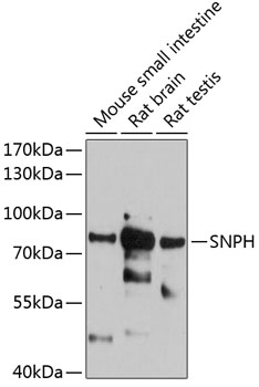
Anti-Mouse/Rat SNPH Antibody144-12300
ApplicationsWestern Blot, ImmunoHistoChemistry
ReactivityHuman, Mouse, Rat
TargetSNPH
- SizePrice
Product group Antibodies
Syntaphilin (N-terminal region) AntibodyBSM-70691M
ApplicationsWestern Blot
ReactivityHuman, Mouse, Rat
TargetSNPH
- SizePrice
Product group Antibodies
SNPH AntibodyCSB-PA022317LA01HU
ApplicationsELISA
ReactivityHuman
TargetSNPH
- SizePrice
Product group Antibodies

SNPH AntibodyLS-C747431
ApplicationsWestern Blot, ImmunoHistoChemistry
ReactivityHuman, Mouse, Rat
TargetSNPH
- SizePrice
Product group Antibodies
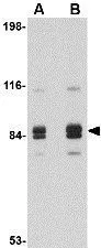
Anti-SNPH AntibodyA81146
ApplicationsWestern Blot, ImmunoHistoChemistry
ReactivityHuman, Mouse, Rat
- SizePrice
Product group Antibodies
Syntaphilin antibodyGTX85393
ApplicationsWestern Blot, ELISA, ImmunoHistoChemistry, ImmunoHistoChemistry Paraffin
ReactivityHuman, Mouse, Rat
TargetSNPH
- SizePrice

Product group Antibodies
Anti-SNPH Antibody Picoband(r)A08882-1-CARRIER-FREE
ApplicationsWestern Blot, ELISA, ImmunoHistoChemistry
ReactivityHuman, Mouse, Rat
TargetSNPH
- SizePrice