

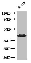
Anti-SPDEF Antibody
144-66214
ApplicationsWestern Blot
Product group Antibodies
ReactivityHuman
TargetSPDEF
Overview
- SupplierRayBiotech
- Product NameAnti-SPDEF Antibody
- Delivery Days Customer16
- ApplicationsWestern Blot
- CertificationResearch Use Only
- ConjugateUnconjugated
- Gene ID25803
- Target nameSPDEF
- Target descriptionSAM pointed domain containing ETS transcription factor
- Target synonymsPDEF, bA375E1.3, SAM pointed domain-containing Ets transcription factor, prostate epithelium-specific Ets transcription factor, prostate-derived Ets factor, prostate-specific Ets
- HostRabbit
- IsotypeIgG
- Protein IDO95238
- Protein NameSAM pointed domain-containing Ets transcription factor
- Scientific DescriptionSPDEF pAb
- ReactivityHuman
- Storage Instruction-20°C
- UNSPSC12352203
Related products
Product group Antibodies
SPDEF AntibodyCSB-PA022518LA01HU
ApplicationsWestern Blot, ELISA, ImmunoHistoChemistry
ReactivityHuman, Mouse
TargetSPDEF
- SizePrice
Product group Antibodies
Spdef Polyclonal AntibodyCAC07726
ApplicationsWestern Blot, ELISA, ImmunoHistoChemistry
ReactivityMouse
TargetSPDEF
- SizePrice
Product group Antibodies
Anti-SPDEF AntibodyA31572
ApplicationsWestern Blot, ImmunoHistoChemistry
ReactivityHuman
- SizePrice

Product group Antibodies
Anti-SPDEF AntibodyHPA055707
ApplicationsImmunoCytoChemistry
ReactivityHuman
TargetSPDEF
- SizePrice

Product group Antibodies
Anti-PSE/SPDEF Antibody Picoband(r)A04625-1-CARRIER-FREE
ApplicationsFlow Cytometry, ImmunoFluorescence, Western Blot, ELISA, ImmunoCytoChemistry
ReactivityHuman
TargetSPDEF
- SizePrice
Product group Antibodies

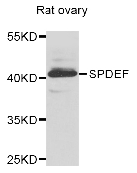
PDEF / SPDEF AntibodyLS-C400961
ApplicationsELISA, ImmunoHistoChemistry
ReactivityHuman, Mouse
TargetSPDEF
- SizePrice
Product group Antibodies
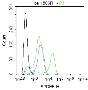
SPDEF Polyclonal AntibodyBS-1866R
ApplicationsFlow Cytometry, ImmunoFluorescence, ELISA, ImmunoCytoChemistry, ImmunoHistoChemistry, ImmunoHistoChemistry Frozen, ImmunoHistoChemistry Paraffin
ReactivityBovine, Canine, Chicken, Equine, Human, Mouse, Rabbit, Rat
TargetSPDEF
- SizePrice