


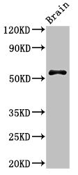
Immunohistochemical staining of human gallbladder shows strong cytoplasmic positivity in glandular cells.
Immunohistochemical staining of human gallbladder shows strong cytoplasmic positivity in glandular cells.
Anti-SRP54 Antibody
HPA048977
ApplicationsImmunoHistoChemistry
Product group Antibodies
ReactivityHuman
TargetSRP54
Overview
- SupplierAtlas Antibodies
- Product NameAnti-SRP54 Antibody
- Delivery Days Customer4
- ApplicationsImmunoHistoChemistry
- CertificationResearch Use Only
- ClonalityPolyclonal
- ConjugateUnconjugated
- Gene ID6729
- Target nameSRP54
- Target descriptionsignal recognition particle 54
- Target synonymsSCN8, signal recognition particle subunit SRP54, signal recognition particle 54 kDa protein, signal recognition particle 54kD, signal recognition particle 54kDa
- HostRabbit
- IsotypeIgG
- Protein IDP61011
- Protein NameSignal recognition particle subunit SRP54
- Scientific DescriptionRecombinant Protein Epitope Signature Tag (PrEST) antigen sequence
- ReactivityHuman
- Storage Instruction-20°C,2°C to 8°C
- UNSPSC41116161
Datasheet
MSDS
Related products
Product group Antibodies
Anti-SRP54 AntibodyA11722
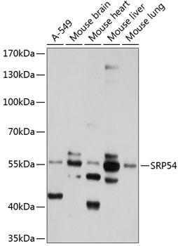
ApplicationsWestern Blot
ReactivityHuman, Mouse
- SizePrice

Product group Antibodies
Anti-SRP54 Antibody Picoband(r)A06189-1-CARRIER-FREE
ApplicationsFlow Cytometry, ImmunoFluorescence, Western Blot, ELISA, ImmunoCytoChemistry
ReactivityHuman, Mouse, Rat
TargetSRP54
- SizePrice
Product group Antibodies
Anti-SRP54 Antibody144-65770
ApplicationsWestern Blot
ReactivityHuman, Mouse
TargetSRP54
- SizePrice
Product group Antibodies

SRP54 AntibodyLS-C770878
ApplicationsELISA, ImmunoHistoChemistry
ReactivityHuman, Mouse, Rat
TargetSRP54
- SizePrice
Product group Antibodies
SRP54 Recombinant AntibodyBSM-61994R
ApplicationsFlow Cytometry, ImmunoFluorescence, ImmunoPrecipitation, Western Blot, ImmunoHistoChemistry, ImmunoHistoChemistry Frozen, ImmunoHistoChemistry Paraffin
ReactivityHuman, Mouse, Rat
TargetSRP54
- SizePrice
Product group Antibodies
SRP54 Polyclonal AntibodyCAC13942
ApplicationsImmunoFluorescence, Western Blot, ELISA
ReactivityMouse
TargetSRP54
- SizePrice
Product group Antibodies
SRP54 AntibodyCSB-PA022675LA01HU
ApplicationsImmunoFluorescence, Western Blot, ELISA
ReactivityHuman, Mouse
TargetSRP54
- SizePrice

Product group Antibodies
Anti-SRP54 AntibodyHPA062044
ApplicationsWestern Blot, ImmunoCytoChemistry
ReactivityHuman
TargetSRP54
- SizePrice