

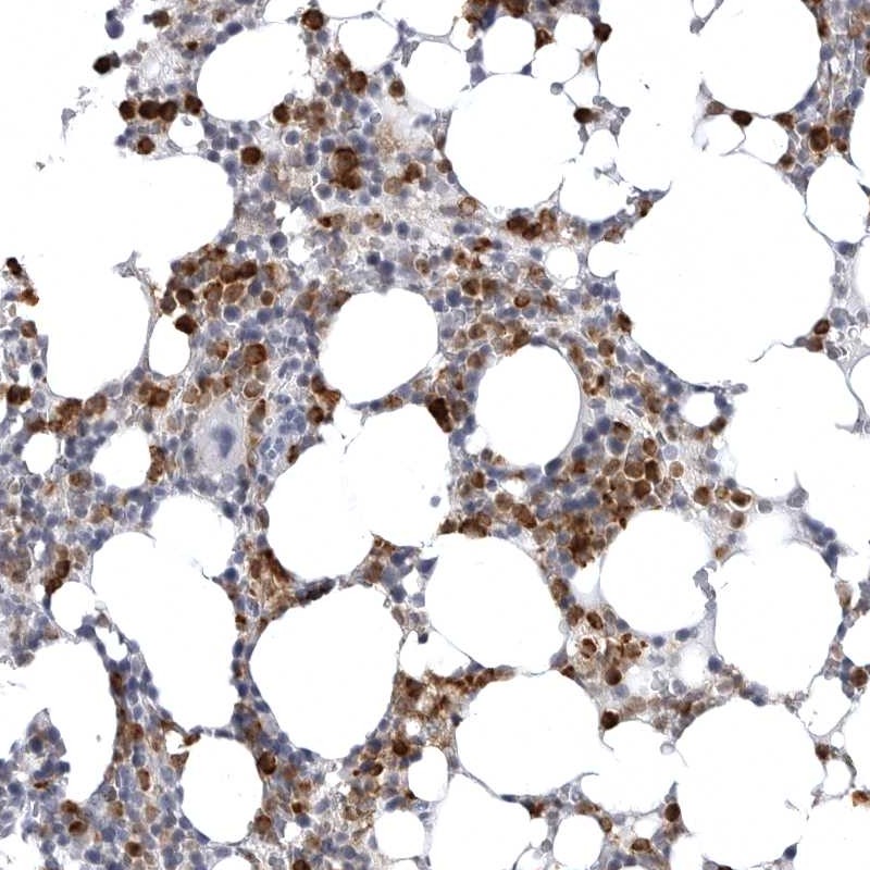
Immunohistochemical staining of human bone marrow shows strong cytoplasmic positivity in subsets of cells.
Immunohistochemical staining of human bone marrow shows strong cytoplasmic positivity in subsets of cells.
Anti-STAP2 Antibody
HPA027761
ApplicationsImmunoHistoChemistry
Product group Antibodies
ReactivityHuman
TargetSTAP2
Overview
- SupplierAtlas Antibodies
- Product NameAnti-STAP2 Antibody
- Delivery Days Customer4
- ApplicationsImmunoHistoChemistry
- CertificationResearch Use Only
- ClonalityPolyclonal
- ConjugateUnconjugated
- Gene ID55620
- Target nameSTAP2
- Target descriptionsignal transducing adaptor family member 2
- Target synonymsBKS, signal-transducing adaptor protein 2, BRK substrate, breast tumor kinase substrate, brk kinase substrate
- HostRabbit
- IsotypeIgG
- Protein IDQ9UGK3
- Protein NameSignal-transducing adaptor protein 2
- Scientific DescriptionRecombinant Protein Epitope Signature Tag (PrEST) antigen sequence
- ReactivityHuman
- Storage Instruction-20°C,2°C to 8°C
- UNSPSC41116161
Datasheet
MSDS
Related products
Product group Antibodies
Anti-STAP2 AntibodyA28474
ApplicationsWestern Blot
ReactivityHuman, Mouse, Rat
- SizePrice

Product group Antibodies
Anti-STAP2 Antibody Picoband(r)A08413-3-CARRIER-FREE
ApplicationsFlow Cytometry, Western Blot, ELISA, ImmunoHistoChemistry
ReactivityHuman
TargetSTAP2
- SizePrice
Product group Antibodies
Anti-STAP2 Antibody144-64131
ApplicationsWestern Blot
ReactivityHuman, Mouse
TargetSTAP2
- SizePrice
Product group Antibodies
STAP2 Polyclonal AntibodyBS-13667R
ApplicationsImmunoFluorescence, Western Blot, ELISA, ImmunoCytoChemistry, ImmunoHistoChemistry, ImmunoHistoChemistry Frozen, ImmunoHistoChemistry Paraffin
ReactivityHuman
TargetSTAP2
- SizePrice
Product group Antibodies

ApplicationsWestern Blot, ELISA, ImmunoHistoChemistry
ReactivityHuman
TargetSTAP2
- SizePrice
Product group Antibodies
Anti-STAP2 AntibodyHPA002375
ApplicationsWestern Blot, ImmunoHistoChemistry
ReactivityHuman
TargetSTAP2
- SizePrice
Product group Antibodies
STAP2 antibody [C3], C-termGTX106287
ApplicationsImmunoFluorescence, Western Blot, ImmunoCytoChemistry, ImmunoHistoChemistry, ImmunoHistoChemistry Paraffin
ReactivityHuman
TargetSTAP2
- SizePrice
Product group Antibodies

STAP2 Antibody (Internal)LS-C353484
ApplicationsWestern Blot
ReactivityHuman, Mouse, Rat
TargetSTAP2
- SizePrice