


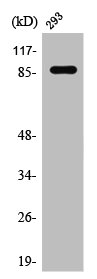
Figure 1. Western blot analysis of STAT1 using anti-STAT1 antibody (M00036). Electrophoresis was performed on a 5-20% SDS-PAGE gel at 70V (Stacking gel) / 90V (Resolving gel) for 2-3 hours. The sample well of each lane was loaded with 30 ug of sample under reducing conditions. Lane 1: human SiHa whole cell lysates, Lane 2: human A549 whole cell lysates, Lane 3: human Caco-2 whole cell lysates, Lane 4: human PC-3 whole cell lysates, Lane 5: human A431 whole cell lysates, Lane 6: human MCF-7 whole cell lysates, Lane 7: human RT4 whole cell lysates, Lane 8: human HEL whole cell lysates. After electrophoresis, proteins were transferred to a nitrocellulose membrane at 150 mA for 50-90 minutes. Blocked the membrane with 5% non-fat milk/TBS for 1.5 hour at RT. The membrane was incubated with rabbit anti-STAT1 antigen affinity purified monoclonal antibody (Catalog # M00036) at 1:5000 overnight at 4°C, then washed with TBS-0.1%Tween 3 times with 5 minutes each and probed with a goat anti-rabbit IgG-HRP secondary antibody at a dilution of 1:500 for 1.5 hour at RT. The signal is developed using an Enhanced Chemiluminescent detection (ECL) kit (Catalog # EK1002) with Tanon 5200 system. A specific band was detected for STAT1 at approximately 91 kDa. The expected band size for STAT1 is at 91 kDa.
Figure 1. Western blot analysis of STAT1 using anti-STAT1 antibody (M00036). Electrophoresis was performed on a 5-20% SDS-PAGE gel at 70V (Stacking gel) / 90V (Resolving gel) for 2-3 hours. The sample well of each lane was loaded with 30 ug of sample under reducing conditions. Lane 1: human SiHa whole cell lysates, Lane 2: human A549 whole cell lysates, Lane 3: human Caco-2 whole cell lysates, Lane 4: human PC-3 whole cell lysates, Lane 5: human A431 whole cell lysates, Lane 6: human MCF-7 whole cell lysates, Lane 7: human RT4 whole cell lysates, Lane 8: human HEL whole cell lysates. After electrophoresis, proteins were transferred to a nitrocellulose membrane at 150 mA for 50-90 minutes. Blocked the membrane with 5% non-fat milk/TBS for 1.5 hour at RT. The membrane was incubated with rabbit anti-STAT1 antigen affinity purified monoclonal antibody (Catalog # M00036) at 1:5000 overnight at 4°C, then washed with TBS-0.1%Tween 3 times with 5 minutes each and probed with a goat anti-rabbit IgG-HRP secondary antibody at a dilution of 1:500 for 1.5 hour at RT. The signal is developed using an Enhanced Chemiluminescent detection (ECL) kit (Catalog # EK1002) with Tanon 5200 system. A specific band was detected for STAT1 at approximately 91 kDa. The expected band size for STAT1 is at 91 kDa.
Anti-STAT1 alpha Rabbit Monoclonal Antibody
M00036
ApplicationsFlow Cytometry, ImmunoFluorescence, ImmunoPrecipitation, Western Blot, ImmunoCytoChemistry, ImmunoHistoChemistry
Product group Antibodies
ReactivityHuman
TargetSTAT1
Overview
- SupplierBoster Bio
- Product NameAnti-STAT1 alpha Rabbit Monoclonal Antibody
- Delivery Days Customer9
- ApplicationsFlow Cytometry, ImmunoFluorescence, ImmunoPrecipitation, Western Blot, ImmunoCytoChemistry, ImmunoHistoChemistry
- CertificationResearch Use Only
- ClonalityMonoclonal
- Clone IDHBG-19
- Gene ID6772
- Target nameSTAT1
- Target descriptionsignal transducer and activator of transcription 1
- Target synonymsCANDF7, IMD31A, IMD31B, IMD31C, ISGF-3, STAT91, signal transducer and activator of transcription 1-alpha/beta, signal transducer and activator of transcription 1, 91kD, signal transducer and activator of transcription 1, 91kDa, transcription factor ISGF-3 components p91/p84
- HostRabbit
- IsotypeIgG
- Protein IDP42224
- Protein NameSignal transducer and activator of transcription 1-alpha/beta
- Scientific DescriptionBoster Bio Anti-STAT1 alpha Rabbit Monoclonal Antibody catalog # M00036. Tested in WB, IHC, ICC/IF, IP, Flow Cytometry applications. This antibody reacts with Human.
- ReactivityHuman
- Storage Instruction-20°C
- UNSPSC12352203
References
- Guan YQ, Li Z, Liu JM. Death signal transduction induced by co-immobilized TNF-α plus IFN-γ and the development of polymeric anti-cancer drugs. Biomaterials. 2010,31(34):9074-85. doi: 10.1016/j.biomaterials.2010.08.044Read this paper
Datasheet
MSDS
Related products
Product group Antibodies
Anti-STAT1 AntibodyA100762
ApplicationsWestern Blot, ELISA
ReactivityHuman
- SizePrice
Product group Antibodies
Anti-P-STAT1 Antibody130-10608
ApplicationsELISA
ReactivityHuman
TargetSTAT1
- SizePrice
Product group Antibodies

Anti-STAT1 [AbAb1-STAT1]AB04098-10.0
ApplicationsELISA
ReactivityHuman
TargetSTAT1
- SizePrice
Product group Antibodies

STAT1 Antibody (Tyr701)LS-C769543
ApplicationsImmunoFluorescence, Western Blot, ELISA, ImmunoHistoChemistry, ImmunoHistoChemistry Paraffin
ReactivityHuman, Monkey, Mouse, Rat
TargetSTAT1
- SizePrice

Product group Antibodies
Anti-STAT1 Antibody Picoband(r)A00036-2-CARRIER-FREE
ApplicationsFlow Cytometry, ImmunoFluorescence, Western Blot, ImmunoCytoChemistry, ImmunoHistoChemistry
ReactivityHuman
TargetSTAT1
- SizePrice
Product group Antibodies
References
STAT1 Polyclonal AntibodyBS-1317R
ApplicationsImmunoFluorescence, Western Blot, ELISA, ImmunoCytoChemistry, ImmunoHistoChemistry, ImmunoHistoChemistry Frozen, ImmunoHistoChemistry Paraffin
ReactivityBovine, Canine, Equine, Human, Mouse, Porcine, Rat, Sheep
TargetSTAT1
- SizePrice
Product group Antibodies
STAT1 AntibodyCSB-PA004167
ApplicationsWestern Blot, ELISA, ImmunoHistoChemistry
ReactivityHuman, Mouse, Rat
TargetSTAT1
- SizePrice
Product group Antibodies
STAT1 Polyclonal AntibodyCAC15929
ApplicationsImmunoPrecipitation, Western Blot, ImmunoHistoChemistry
ReactivityMouse, Primate, Rat
TargetSTAT1
- SizePrice
Product group Antibodies
STAT1 (phospho Ser727) antibodyGTX132507
ApplicationsWestern Blot
ReactivityHuman
TargetSTAT1
- SizePrice