


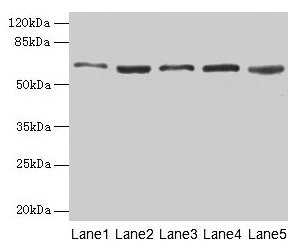
Figure 1. Western blot analysis of STIP1 using anti-STIP1 antibody (PB9896). Electrophoresis was performed on a 5-20% SDS-PAGE gel at 70V (Stacking gel) / 90V (Resolving gel) for 2-3 hours. The sample well of each lane was loaded with 30 ug of sample under reducing conditions. Lane 1: human Hela whole cell lysates, Lane 2: human COLO320 whole cell lysates, Lane 3: human MCF-7 whole cell lysates, Lane 4: rat C6 whole cell lysates, Lane 5: rat RH35 whole cell lysates, Lane 6: mouse brain tissue lysates, Lane 7: mouse liver tissue lysates, Lane 8: mouse Neuro-2a whole cell lysates, Lane 9: mouse HEPA1-6 whole cell lysates. After electrophoresis, proteins were transferred to a nitrocellulose membrane at 150 mA for 50-90 minutes. Blocked the membrane with 5% non-fat milk/TBS for 1.5 hour at RT. The membrane was incubated with rabbit anti-STIP1 antigen affinity purified polyclonal antibody (Catalog # PB9896) at 0.5 microg/mL overnight at 4°C, then washed with TBS-0.1%Tween 3 times with 5 minutes each and probed with a goat anti-rabbit IgG-HRP secondary antibody at a dilution of 1:5000 for 1.5 hour at RT. The signal is developed using an Enhanced Chemiluminescent detection (ECL) kit (Catalog # EK1002) with Tanon 5200 system. A specific band was detected for STIP1 at approximately 63 kDa. The expected band size for STIP1 is at 63 kDa.
Figure 1. Western blot analysis of STIP1 using anti-STIP1 antibody (PB9896). Electrophoresis was performed on a 5-20% SDS-PAGE gel at 70V (Stacking gel) / 90V (Resolving gel) for 2-3 hours. The sample well of each lane was loaded with 30 ug of sample under reducing conditions. Lane 1: human Hela whole cell lysates, Lane 2: human COLO320 whole cell lysates, Lane 3: human MCF-7 whole cell lysates, Lane 4: rat C6 whole cell lysates, Lane 5: rat RH35 whole cell lysates, Lane 6: mouse brain tissue lysates, Lane 7: mouse liver tissue lysates, Lane 8: mouse Neuro-2a whole cell lysates, Lane 9: mouse HEPA1-6 whole cell lysates. After electrophoresis, proteins were transferred to a nitrocellulose membrane at 150 mA for 50-90 minutes. Blocked the membrane with 5% non-fat milk/TBS for 1.5 hour at RT. The membrane was incubated with rabbit anti-STIP1 antigen affinity purified polyclonal antibody (Catalog # PB9896) at 0.5 microg/mL overnight at 4°C, then washed with TBS-0.1%Tween 3 times with 5 minutes each and probed with a goat anti-rabbit IgG-HRP secondary antibody at a dilution of 1:5000 for 1.5 hour at RT. The signal is developed using an Enhanced Chemiluminescent detection (ECL) kit (Catalog # EK1002) with Tanon 5200 system. A specific band was detected for STIP1 at approximately 63 kDa. The expected band size for STIP1 is at 63 kDa.
Anti-STIP1 Antibody Picoband(r)
PB9896-CARRIER-FREE
ApplicationsWestern Blot
Product group Antibodies
ReactivityBovine, Canine, Equine, Human, Monkey, Mouse, Rabbit, Rat
TargetSTIP1
Overview
- SupplierBoster Bio
- Product NameAnti-STIP1 Antibody Picoband(r)
- Delivery Days Customer9
- Application Supplier NoteTested Species: In-house tested species with positive results. Other applications have not been tested. Optimal dilutions should be determined by end users.
- ApplicationsWestern Blot
- CertificationResearch Use Only
- ClonalityPolyclonal
- Concentration500 ug/ml
- Gene ID10963
- Target nameSTIP1
- Target descriptionstress induced phosphoprotein 1
- Target synonymsHEL-S-94n, HOP, IEF-SSP-3521, P60, STI1, STI1L, stress-induced-phosphoprotein 1, Hsp70/Hsp90-organizing protein, NY-REN-11 antigen, epididymis secretory sperm binding protein Li 94n, hsc70/Hsp90-organizing protein, renal carcinoma antigen NY-REN-11, transformation-sensitive protein IEF SSP 3521
- HostRabbit
- IsotypeIgG
- Protein IDP31948
- Protein NameStress-induced-phosphoprotein 1
- Scientific DescriptionBoster Bio Anti-STIP1 Antibody Picoband® catalog # PB9896. Tested in WB applications. This antibody reacts with Human, Mouse, Rat. The brand Picoband indicates this is a premium antibody that guarantees superior quality, high affinity, and strong signals with minimal background in Western blot applications. Only our best-performing antibodies are designated as Picoband, ensuring unmatched performance.
- ReactivityBovine, Canine, Equine, Human, Monkey, Mouse, Rabbit, Rat
- Storage Instruction-20°C,2°C to 8°C
- UNSPSC12352203
Related products
Product group Antibodies
STIP1 AntibodyCSB-PA022831HA01HU
ApplicationsWestern Blot, ELISA, ImmunoHistoChemistry
ReactivityHuman, Mouse, Rat
TargetSTIP1
- SizePrice
Product group Antibodies
Anti-STIP1 Antibody144-01219
ApplicationsImmunoFluorescence, Western Blot, ImmunoHistoChemistry
ReactivityHuman, Monkey, Mouse
TargetSTIP1
- SizePrice

Product group Antibodies
Anti-STIP1 AntibodyA29510
ApplicationsImmunoFluorescence, ImmunoPrecipitation, Western Blot, ImmunoHistoChemistry, Other Application
ReactivityHuman, Mouse, Rat
- SizePrice
Product group Antibodies

STI1 / STIP1 AntibodyLS-C749119
ApplicationsImmunoFluorescence, ImmunoPrecipitation, Western Blot
ReactivityHuman, Monkey, Mouse
TargetSTIP1
- SizePrice
Product group Antibodies
Anti-STIP1 AntibodyHPA039291
ApplicationsWestern Blot, ImmunoCytoChemistry, ImmunoHistoChemistry
ReactivityHuman
TargetSTIP1
- SizePrice
Product group Antibodies
STIP1 Recombinant AntibodyBSM-60759R
ApplicationsFlow Cytometry, ImmunoFluorescence, Western Blot, ImmunoCytoChemistry, ImmunoHistoChemistry, ImmunoHistoChemistry Frozen, ImmunoHistoChemistry Paraffin
ReactivityHuman, Mouse, Rat
TargetSTIP1
- SizePrice
Product group Antibodies
STIP1 antibodyGTX103019
ApplicationsImmunoFluorescence, Western Blot, ImmunoCytoChemistry, ImmunoHistoChemistry, ImmunoHistoChemistry Paraffin
ReactivityHuman
TargetSTIP1
- SizePrice