

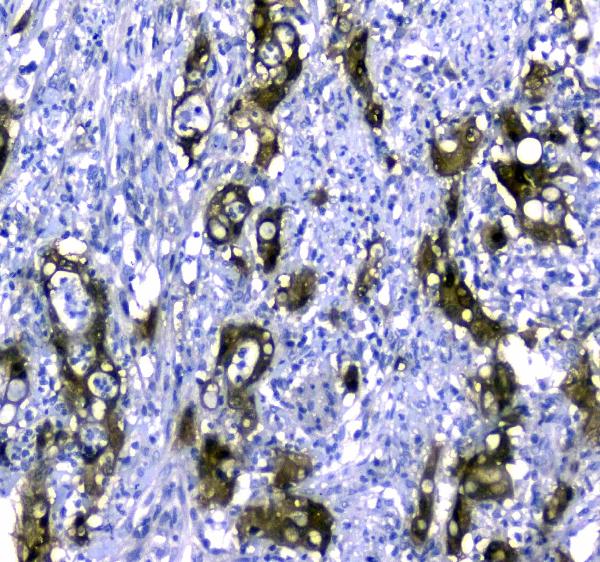
Figure 2. IHC analysis of STUB1 using anti-STUB1 antibody (A01236-1). STUB1 was detected in paraffin-embedded section of human colon cancer tissue. Heat mediated antigen retrieval was performed in citrate buffer (pH6, epitope retrieval solution) for 20 mins. The tissue section was blocked with 10% goat serum. The tissue section was then incubated with 1microg/ml rabbit anti-STUB1 Antibody (A01236-1) overnight at 4°C. Biotinylated goat anti-rabbit IgG was used as secondary antibody and incubated for 30 minutes at 37°C. The tissue section was developed using Strepavidin-Biotin-Complex (SABC)(Catalog # SA1022) with DAB as the chromogen.
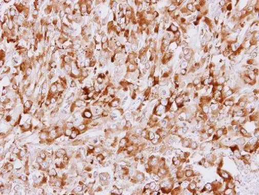
. STUB1 was detected in paraffin-embedded section of human lung cancer tissue. Heat mediated antigen retrieval was performed in citrate buffer (pH6, epitope retrieval solution) for 20 mins. The tissue section was blocked with 10% goat serum. The tissue section was then incubated with 1microg/ml rabbit anti-STUB1 Antibody (A01236-1) overnight at 4°C. Biotinylated goat anti-rabbit IgG was used as secondary antibody and incubated for 30 minutes at 37°C. The tissue section was developed using Strepavidin-Biotin-Complex (SABC)(Catalog # SA1022) with DAB as the chromogen.")
. STUB1 was detected in paraffin-embedded section of human mammary cancer tissue. Heat mediated antigen retrieval was performed in citrate buffer (pH6, epitope retrieval solution) for 20 mins. The tissue section was blocked with 10% goat serum. The tissue section was then incubated with 1microg/ml rabbit anti-STUB1 Antibody (A01236-1) overnight at 4°C. Biotinylated goat anti-rabbit IgG was used as secondary antibody and incubated for 30 minutes at 37°C. The tissue section was developed using Strepavidin-Biotin-Complex (SABC)(Catalog # SA1022) with DAB as the chromogen.")
. STUB1 was detected in immunocytochemical section of MCF7 cells. Enzyme antigen retrieval was performed using IHC enzyme antigen retrieval reagent (AR0022) for 15 mins. The cells were blocked with 10% goat serum. And then incubated with 2microg/mL rabbit anti-STUB1 Antibody (A01236-1) overnight at 4°C. DyLight®594 Conjugated Goat Anti-Rabbit IgG (BA1142) was used as secondary antibody at 1:100 dilution and incubated for 30 minutes at 37°C. The section was counterstained with DAPI. Visualize using a fluorescence microscope and filter sets appropriate for the label used.")
. Overlay histogram showing A549 cells stained with A01236-1 (Blue line). To facilitate intracellular staining, cells were fixed with 4% paraformaldehyde and permeabilized with permeabilization buffer. The cells were blocked with 10% normal goat serum. And then incubated with rabbit anti-STUB1 Antibody (A01236-1,1microg/1x106 cells) for 30 min at 20°C. DyLight®488 conjugated goat anti-rabbit IgG (BA1127, 5-10microg/1x106 cells) was used as secondary antibody for 30 minutes at 20°C. Isotype control antibody (Green line) was rabbit IgG (1microg/1x106) used under the same conditions. Unlabelled sample without incubation with primary antibody and secondary antibody (Red line) was used as a blank control.")
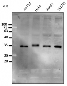
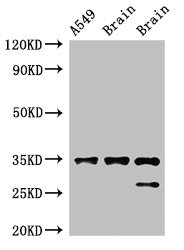
. Electrophoresis was performed on a 5-20% SDS-PAGE gel at 70V (Stacking gel) / 90V (Resolving gel) for 2-3 hours. The sample well of each lane was loaded with 30 ug of sample under reducing conditions. Lane 1: human 293T whole cell lysates, Lane 2: human MCF-7 whole cell lysates, Lane 3: human Hela whole cell lysates, Lane 4: human THP-1 whole cell lysates, Lane 5: rat pancreas tissue lysates, Lane 6: mouse pancreas tissue lysates. After electrophoresis, proteins were transferred to a nitrocellulose membrane at 150 mA for 50-90 minutes. Blocked the membrane with 5% non-fat milk/TBS for 1.5 hour at RT. The membrane was incubated with rabbit anti-STUB1 antigen affinity purified polyclonal antibody (Catalog # A01236-1) at 0.5 microg/mL overnight at 4°C, then washed with TBS-0.1%Tween 3 times with 5 minutes each and probed with a goat anti-rabbit IgG-HRP secondary antibody at a dilution of 1:5000 for 1.5 hour at RT. The signal is developed using an Enhanced Chemiluminescent detection (ECL) kit (Catalog # EK1002) with Tanon 5200 system. A specific band was detected for STUB1 at approximately 35 kDa. The expected band size for STUB1 is at 35 kDa.")
Figure 2. IHC analysis of STUB1 using anti-STUB1 antibody (A01236-1). STUB1 was detected in paraffin-embedded section of human colon cancer tissue. Heat mediated antigen retrieval was performed in citrate buffer (pH6, epitope retrieval solution) for 20 mins. The tissue section was blocked with 10% goat serum. The tissue section was then incubated with 1microg/ml rabbit anti-STUB1 Antibody (A01236-1) overnight at 4°C. Biotinylated goat anti-rabbit IgG was used as secondary antibody and incubated for 30 minutes at 37°C. The tissue section was developed using Strepavidin-Biotin-Complex (SABC)(Catalog # SA1022) with DAB as the chromogen.
Anti-STUB1 Antibody Picoband(r)
A01236-1-CARRIER-FREE
ApplicationsFlow Cytometry, ImmunoFluorescence, Western Blot, ELISA, ImmunoCytoChemistry, ImmunoHistoChemistry
Product group Antibodies
ReactivityHuman, Mouse, Rat
TargetSTUB1
Overview
- SupplierBoster Bio
- Product NameAnti-STUB1 Antibody Picoband(r)
- Delivery Days Customer9
- ApplicationsFlow Cytometry, ImmunoFluorescence, Western Blot, ELISA, ImmunoCytoChemistry, ImmunoHistoChemistry
- CertificationResearch Use Only
- ClonalityPolyclonal
- Concentration500 ug/ml
- Gene ID10273
- Target nameSTUB1
- Target descriptionSTIP1 homology and U-box containing protein 1
- Target synonymsCHIP, HSPABP2, NY-CO-7, SCA48, SCAR16, SDCCAG7, UBOX1, E3 ubiquitin-protein ligase CHIP, CLL-associated antigen KW-8, RING-type E3 ubiquitin transferase CHIP, STIP1 homology and U-box containing protein 1, E3 ubiquitin protein ligase, antigen NY-CO-7, carboxy terminus of Hsp70-interacting protein, heat shock protein A binding protein 2 (c-terminal), serologically defined colon cancer antigen 7
- HostRabbit
- IsotypeIgG
- Protein IDQ9UNE7
- Protein NameE3 ubiquitin-protein ligase CHIP
- Scientific DescriptionBoster Bio Anti-STUB1 Antibody Picoband® catalog # A01236-1. Tested in ELISA, Flow Cytometry, IF, IHC, ICC, WB applications. This antibody reacts with Human, Mouse, Rat. The brand Picoband indicates this is a premium antibody that guarantees superior quality, high affinity, and strong signals with minimal background in Western blot applications. Only our best-performing antibodies are designated as Picoband, ensuring unmatched performance.
- ReactivityHuman, Mouse, Rat
- Storage Instruction-20°C,2°C to 8°C
- UNSPSC12352203
Related products
Product group Antibodies
Anti-STUB1 AntibodyA121663
ApplicationsWestern Blot
ReactivityCanine, Human, Monkey, Mouse, Rat
- SizePrice
Product group Antibodies
Anti-STUB1 Antibody144-11751
ApplicationsWestern Blot
ReactivityHuman, Mouse, Rat
TargetSTUB1
- SizePrice
Product group Antibodies

STUB1 / CHIP AntibodyLS-C747118
ApplicationsWestern Blot
ReactivityHuman, Mouse, Rat
TargetSTUB1
- SizePrice
Product group Antibodies
ApplicationsFlow Cytometry, Western Blot, ImmunoCytoChemistry
ReactivityHuman, Mouse, Rat
TargetSTUB1
- SizePrice
Product group Antibodies

Goat anti-STUB1 / CHIPEB05327
ApplicationsWestern Blot, ELISA, ImmunoHistoChemistry
ReactivityBovine, Canine, Human, Mouse, Rat
TargetSTUB1
- SizePrice
Product group Antibodies
STUB1 AntibodyCSB-PA892480LA01HU
ApplicationsImmunoFluorescence, Western Blot, ELISA, ImmunoHistoChemistry
ReactivityHuman, Mouse, Rat
TargetSTUB1
- SizePrice
Product group Antibodies
Stub1 Polyclonal AntibodyCAC07955
ApplicationsImmunoFluorescence, Western Blot, ELISA, ImmunoHistoChemistry
ReactivityMouse, Rat
TargetSTUB1
- SizePrice

Product group Antibodies
Anti-STUB1 AntibodyHPA041222
ApplicationsWestern Blot, ImmunoHistoChemistry
ReactivityHuman
TargetSTUB1
- SizePrice
Product group Antibodies
STUB1 antibody [C1C3]GTX109676
ApplicationsImmunoFluorescence, Western Blot, ImmunoCytoChemistry, ImmunoHistoChemistry, ImmunoHistoChemistry Paraffin
ReactivityHuman, Mouse
TargetSTUB1
- SizePrice
Product group Antibodies
Anti-STUB1Y058159
ApplicationsWestern Blot, ELISA
ReactivityHuman, Mouse, Rat
- SizePrice