


Immunohistochemical staining of human testis shows strong cytoplasmic and nuclear positivity in cells in seminiferous ducts.
Immunohistochemical staining of human testis shows strong cytoplasmic and nuclear positivity in cells in seminiferous ducts.
Anti-SUGT1 Antibody
HPA069215
ApplicationsImmunoHistoChemistry
Product group Antibodies
ReactivityHuman
TargetSUGT1
Overview
- SupplierAtlas Antibodies
- Product NameAnti-SUGT1 Antibody
- Delivery Days Customer12
- ApplicationsImmunoHistoChemistry
- CertificationResearch Use Only
- ClonalityPolyclonal
- ConjugateUnconjugated
- Gene ID10910
- Target nameSUGT1
- Target descriptionSGT1 assembly cochaperone of MIS12 kinetochore complex
- Target synonymsSGT1, protein SGT1 homolog, SGT1 homolog, MIS12 kinetochore complex assembly cochaperone, SGT1, suppressor of G2 allele of SKP1, putative 40-6-3 protein, suppressor of G2 allele of SKP1, S. cerevisiae, homolog of
- HostRabbit
- IsotypeIgG
- Protein IDQ9Y2Z0
- Protein NameProtein SGT1 homolog
- Scientific DescriptionRecombinant Protein Epitope Signature Tag (PrEST) antigen sequence
- ReactivityHuman
- Storage Instruction-20°C,2°C to 8°C
- UNSPSC41116161
MSDS
Related products
Product group Antibodies
Anti-SUGT1 Antibody101-11691
ApplicationsImmunoFluorescence, Western Blot, ELISA
TargetSUGT1
- SizePrice
Product group Antibodies
SUGT1 Polyclonal AntibodyBS-7831R
ApplicationsImmunoFluorescence, Western Blot, ELISA, ImmunoCytoChemistry, ImmunoHistoChemistry, ImmunoHistoChemistry Frozen, ImmunoHistoChemistry Paraffin
ReactivityBovine, Canine, Equine, Human, Mouse, Porcine, Rabbit, Rat, Sheep
TargetSUGT1
- SizePrice
Product group Antibodies
SUGT1 AntibodyCSB-PA022924GA01HU
ApplicationsWestern Blot, ELISA, ImmunoHistoChemistry
ReactivityHuman, Mouse, Rat
TargetSUGT1
- SizePrice
Product group Antibodies

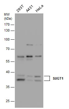
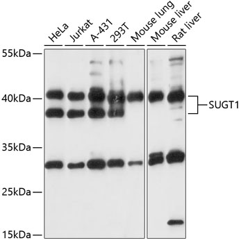
SUGT1 / SGT1 AntibodyLS-C748928
ApplicationsWestern Blot
ReactivityHuman, Mouse, Rat
TargetSUGT1
- SizePrice
Product group Antibodies
SUGT1 antibody [N1C3]GTX105358
ApplicationsImmunoFluorescence, Western Blot, ImmunoCytoChemistry
ReactivityHuman
TargetSUGT1
- SizePrice
Product group Antibodies
Anti-SUGT1 AntibodyA87571
ApplicationsWestern Blot
ReactivityHuman, Mouse, Rat
- SizePrice

Product group Antibodies
Anti-SUGT1 Antibody Picoband(r)A05411-1-CARRIER-FREE
ApplicationsFlow Cytometry, ImmunoFluorescence, Western Blot, ELISA, ImmunoCytoChemistry
ReactivityHuman
TargetSUGT1
- SizePrice