


Immunohistochemical staining of human duodenum shows strong granular cytoplasmic positivity in glandular cells.

Immunohistochemical staining of human duodenum shows strong granular cytoplasmic positivity in glandular cells.
Anti-SYNJ2BP Antibody
HPA000866
ApplicationsWestern Blot, ImmunoCytoChemistry, ImmunoHistoChemistry
Product group Antibodies
ReactivityHuman, Mouse
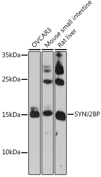
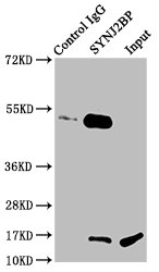
TargetSYNJ2BP
Overview
- SupplierAtlas Antibodies
- Product NameAnti-SYNJ2BP Antibody
- Delivery Days Customer4
- ApplicationsWestern Blot, ImmunoCytoChemistry, ImmunoHistoChemistry
- CertificationResearch Use Only
- ClonalityPolyclonal
- ConjugateUnconjugated
- Gene ID55333
- Target nameSYNJ2BP
- Target descriptionsynaptojanin 2 binding protein
- Target synonymsARIP2, OMP25, synaptojanin-2-binding protein, activin receptor interacting protein 5, mitochondrial outer membrane protein 25
- HostRabbit
- IsotypeIgG
- Protein IDP57105
- Protein NameSynaptojanin-2-binding protein
- Scientific DescriptionRecombinant Protein Epitope Signature Tag (PrEST) antigen sequence
- ReactivityHuman, Mouse
- Storage Instruction-20°C,2°C to 8°C
- UNSPSC41116161
Datasheet
MSDS
Related products
Product group Antibodies
Anti-SYNJ2BP AntibodyA91945
ApplicationsWestern Blot
ReactivityHuman, Mouse, Rat
- SizePrice

Product group Antibodies
Anti-SYNJ2BP Antibody Picoband(r)A11249-1-CARRIER-FREE
ApplicationsWestern Blot, ELISA
ReactivityHuman, Mouse, Rat
TargetSYNJ2BP
- SizePrice
Product group Antibodies

SYNJ2BP / OMP25 AntibodyLS-C750440
ApplicationsWestern Blot
ReactivityHuman, Rat
TargetSYNJ2BP
- SizePrice
Product group Antibodies
Synj2Bp Polyclonal AntibodyCAC08889
ApplicationsImmunoPrecipitation, Western Blot, ELISA, ImmunoHistoChemistry
ReactivityRat
TargetSYNJ2BP
- SizePrice
Product group Antibodies
SYNJ2BP AntibodyCSB-PA023018LA01HU
ApplicationsImmunoPrecipitation, Western Blot, ELISA, ImmunoHistoChemistry
ReactivityHuman, Rat
TargetSYNJ2BP
- SizePrice