


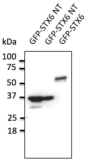
Figure 1. Western blot analysis of Syntaxin 6/STX6 using anti-Syntaxin 6/STX6 antibody (A06586-1). Electrophoresis was performed on a 5-20% SDS-PAGE gel at 70V (Stacking gel) / 90V (Resolving gel) for 2-3 hours. The sample well of each lane was loaded with 30 ug of sample under reducing conditions. Lane 1: human RT4 whole cell lysates, Lane 2: human Hacat whole cell lysates, Lane 3: human SH-SY5Y whole cell lysates, Lane 4: human T-47D whole cell lysates, Lane 5: rat brain tissue lysates, Lane 6: rat PC-12 whole cell lysates, Lane 7: mouse brain tissue lysates, Lane 8: mouse Neuro-2a whole cell lysates. After electrophoresis, proteins were transferred to a nitrocellulose membrane at 150 mA for 50-90 minutes. Blocked the membrane with 5% non-fat milk/TBS for 1.5 hour at RT. The membrane was incubated with rabbit anti-Syntaxin 6/STX6 antigen affinity purified polyclonal antibody (Catalog # A06586-1) at 0.5 microg/mL overnight at 4°C, then washed with TBS-0.1%Tween 3 times with 5 minutes each and probed with a goat anti-rabbit IgG-HRP secondary antibody at a dilution of 1:5000 for 1.5 hour at RT. The signal is developed using an Enhanced Chemiluminescent detection (ECL) kit (Catalog # EK1002) with Tanon 5200 system. A specific band was detected for Syntaxin 6/STX6 at approximately 29 kDa. The expected band size for Syntaxin 6/STX6 is at 29 kDa.
 and anti-Tubulin beta antibody (M05613-4). CAT2/SLC7A2 and Tubulin beta was detected in an immunocytochemical section of A549 cells. Enzyme antigen retrieval was performed using IHC enzyme antigen retrieval reagent (AR0022) for 15 mins. The cells were blocked with 10% goat serum. And then incubated with 5 microg/mL rabbit anti-CAT2/SLC7A2 Antibody (A06586-1) and mouse anti-Tubulin beta Antibody (M05613-4) overnight at 4°C. Cy3 Conjugated Goat Anti-Rabbit IgG (BA1032) and DyLight®488 Conjugated Goat Anti-Mouse IgG (BA1126) were used as secondary antibody at 1:500 dilution and incubated for 30 minutes at 37°C. Visualize using a fluorescence microscope and filter sets appropriate for the label used.")
. Overlay histogram showing U20S cells stained with A06586-1 (Blue line). To facilitate intracellular staining, cells were fixed with 4% paraformaldehyde and permeabilized with permeabilization buffer. The cells were blocked with 10% normal goat serum. And then incubated with rabbit anti-Syntaxin 6/STX6 Antibody (A06586-1, 1 microg/1x106 cells) for 30 min at 20°C. DyLight®488 conjugated goat anti-rabbit IgG (BA1127, 5-10 microg/1x106 cells) was used as secondary antibody for 30 minutes at 20°C. Isotype control antibody (Green line) was rabbit IgG (1 microg/1x106) used under the same conditions. Unlabelled sample without incubation with primary antibody and secondary antibody (Red line) was used as a blank control.")
Figure 1. Western blot analysis of Syntaxin 6/STX6 using anti-Syntaxin 6/STX6 antibody (A06586-1). Electrophoresis was performed on a 5-20% SDS-PAGE gel at 70V (Stacking gel) / 90V (Resolving gel) for 2-3 hours. The sample well of each lane was loaded with 30 ug of sample under reducing conditions. Lane 1: human RT4 whole cell lysates, Lane 2: human Hacat whole cell lysates, Lane 3: human SH-SY5Y whole cell lysates, Lane 4: human T-47D whole cell lysates, Lane 5: rat brain tissue lysates, Lane 6: rat PC-12 whole cell lysates, Lane 7: mouse brain tissue lysates, Lane 8: mouse Neuro-2a whole cell lysates. After electrophoresis, proteins were transferred to a nitrocellulose membrane at 150 mA for 50-90 minutes. Blocked the membrane with 5% non-fat milk/TBS for 1.5 hour at RT. The membrane was incubated with rabbit anti-Syntaxin 6/STX6 antigen affinity purified polyclonal antibody (Catalog # A06586-1) at 0.5 microg/mL overnight at 4°C, then washed with TBS-0.1%Tween 3 times with 5 minutes each and probed with a goat anti-rabbit IgG-HRP secondary antibody at a dilution of 1:5000 for 1.5 hour at RT. The signal is developed using an Enhanced Chemiluminescent detection (ECL) kit (Catalog # EK1002) with Tanon 5200 system. A specific band was detected for Syntaxin 6/STX6 at approximately 29 kDa. The expected band size for Syntaxin 6/STX6 is at 29 kDa.
Anti-Syntaxin 6/STX6 Antibody Picoband(r)
A06586-1-IFLUOR647
ApplicationsFlow Cytometry, Western Blot, ELISA
Product group Antibodies
ReactivityHuman, Mouse, Rat
TargetSTX6
Overview
- SupplierBoster Bio
- Product NameAnti-Syntaxin 6/STX6 Antibody Picoband(r)
- Delivery Days Customer9
- ApplicationsFlow Cytometry, Western Blot, ELISA
- CertificationResearch Use Only
- ClonalityPolyclonal
- Concentration500 ug/ml
- ConjugateOther Conjugate
- Gene ID10228
- Target nameSTX6
- Target descriptionsyntaxin 6
- Target synonymssyntaxin-6
- HostRabbit
- IsotypeIgG
- Protein IDO43752
- Protein NameSyntaxin-6
- Scientific DescriptionBoster Bio Anti-Syntaxin 6/STX6 Antibody Picoband® catalog # A06586-1. Tested in ELISA, Flow Cytometry, WB applications. This antibody reacts with Human, Mouse, Rat. The brand Picoband indicates this is a premium antibody that guarantees superior quality, high affinity, and strong signals with minimal background in Western blot applications. Only our best-performing antibodies are designated as Picoband, ensuring unmatched performance.
- ReactivityHuman, Mouse, Rat
- Storage Instruction-20°C,2°C to 8°C
- UNSPSC12352203
Related products
Product group Antibodies
STX6 Monoclonal AntibodyBSM-60361M
ApplicationsWestern Blot
ReactivityHuman, Rat
TargetSTX6
- SizePrice
Product group Antibodies
Anti-STX6 AntibodyA121585
ApplicationsWestern Blot
ReactivityCanine, Human, Monkey, Mouse, Rat
- SizePrice
Product group Antibodies

Goat anti-STX6 AntibodyEB07214
ApplicationsWestern Blot, ELISA
ReactivityCanine, Human, Mouse, Rat
TargetSTX6
- SizePrice
Product group Antibodies
References
Syntaxin 6 antibody [N1C3]GTX115375
ApplicationsImmunoFluorescence, Western Blot, ImmunoCytoChemistry, ImmunoHistoChemistry, ImmunoHistoChemistry Paraffin
ReactivityHuman, Mouse, Rat
TargetSTX6
- SizePrice
Product group Antibodies

STX6 / Syntaxin 6 AntibodyLS-C830950
ApplicationsELISA, ImmunoHistoChemistry
ReactivityHuman, Mouse, Rat
TargetSTX6
- SizePrice
Product group Antibodies
Anti-STX6 AntibodyHPA038558
ApplicationsWestern Blot, ImmunoCytoChemistry, ImmunoHistoChemistry
ReactivityHuman
TargetSTX6
- SizePrice
Product group Antibodies
Anti-Syntaxin 6/STX6 Antibody Picoband(r)A06586-1-CARRIER-FREE
ApplicationsFlow Cytometry, Western Blot, ELISA
ReactivityHuman, Mouse, Rat
TargetSTX6
- SizePrice
Product group Antibodies
STX6 AntibodyCSB-PA022902DA01HU
ApplicationsELISA, ImmunoHistoChemistry
ReactivityHuman
TargetSTX6
- SizePrice