


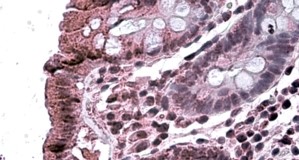
Immunohistochemical staining of human pancreas shows cytoplasmic positivity in exocrine cells.
Immunohistochemical staining of human pancreas shows cytoplasmic positivity in exocrine cells.
Anti-TAB2 Antibody
HPA065436
ApplicationsImmunoHistoChemistry
Product group Antibodies
ReactivityHuman
TargetTAB2
Overview
- SupplierAtlas Antibodies
- Product NameAnti-TAB2 Antibody
- Delivery Days Customer4
- ApplicationsImmunoHistoChemistry
- CertificationResearch Use Only
- ClonalityPolyclonal
- ConjugateUnconjugated
- Gene ID23118
- Target nameTAB2
- Target descriptionTGF-beta activated kinase 1 (MAP3K7) binding protein 2
- Target synonymsCHTD2, MAP3K7IP2, TAB-2, TGF-beta-activated kinase 1 and MAP3K7-binding protein 2, TAK1-binding protein 2, mitogen-activated protein kinase kinase kinase 7-interacting protein 2
- HostRabbit
- IsotypeIgG
- Protein IDQ9NYJ8
- Protein NameTGF-beta-activated kinase 1 and MAP3K7-binding protein 2
- Scientific DescriptionRecombinant Protein Epitope Signature Tag (PrEST) antigen sequence
- ReactivityHuman
- Storage Instruction-20°C,2°C to 8°C
- UNSPSC41116161
Datasheet
MSDS
Related products
Product group Antibodies
TAB2 AntibodyCSB-PA013431GA01HU
ApplicationsImmunoFluorescence, Western Blot, ELISA, ImmunoHistoChemistry
ReactivityHuman, Mouse, Rat
TargetTAB2
- SizePrice
Product group Antibodies
Anti-TAB2 AntibodyA326305
ApplicationsELISA, ImmunoHistoChemistry
ReactivityHuman
- SizePrice

Product group Antibodies
Anti-TAB2 Antibody Picoband(r)A02499-1-CARRIER-FREE
ApplicationsFlow Cytometry, Western Blot, ELISA, ImmunoHistoChemistry
ReactivityHuman
TargetTAB2
- SizePrice
Product group Antibodies

Goat anti-MAP3K7IP2EB09916
ApplicationsELISA, ImmunoHistoChemistry
ReactivityBovine, Canine, Human, Mouse, Rat
TargetTAB2
- SizePrice

Product group Antibodies
Anti-TAB2 AntibodyHPA070137
ApplicationsImmunoHistoChemistry
ReactivityHuman
TargetTAB2
- SizePrice

Product group Antibodies
Anti-TAB2 AntibodyHPA071215
ApplicationsImmunoCytoChemistry
ReactivityHuman
TargetTAB2
- SizePrice
Product group Antibodies

TAB2 AntibodyLS-C498046
ApplicationsWestern Blot
ReactivityHuman, Mouse
TargetTAB2
- SizePrice