


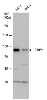
Figure 1. Western blot analysis of TAP1 using anti-TAP1 antibody (PB9823). Electrophoresis was performed on a 5-20% SDS-PAGE gel at 70V (Stacking gel) / 90V (Resolving gel) for 2-3 hours. The sample well of each lane was loaded with 40 ug of sample under reducing conditions. Lane 1: HELA Whole Cell Lysate, Lane 2: HUT Whole Cell Lysate, Lane 3: SW620 Whole Cell Lysate. After electrophoresis, proteins were transferred to a nitrocellulose membrane at 150 mA for 50-90 minutes. Blocked the membrane with 5% non-fat milk/TBS for 1.5 hour at RT. The membrane was incubated with rabbit anti-TAP1 antigen affinity purified polyclonal antibody (Catalog # PB9823) at 0.5 microg/mL overnight at 4°C, then washed with TBS-0.1%Tween 3 times with 5 minutes each and probed with a goat anti-rabbit IgG-HRP secondary antibody at a dilution of 1:5000 for 1.5 hour at RT. The signal is developed using an Enhanced Chemiluminescent detection (ECL) kit (Catalog # EK1002) with Tanon 5200 system. A specific band was detected for TAP1 at approximately 87 kDa. The expected band size for TAP1 is at 87 kDa.
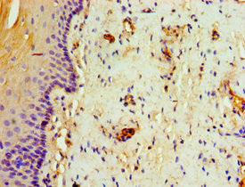
. TAP1 was detected in a paraffin-embedded section of human intestinal cancer tissue. Heat mediated antigen retrieval was performed in EDTA buffer (pH 8.0, epitope retrieval solution). The tissue section was blocked with 10% goat serum. The tissue section was then incubated with 1 microg/ml rabbit anti-TAP1 Antibody (PB9823) overnight at 4°C. Biotinylated goat anti-rabbit IgG was used as secondary antibody and incubated for 30 minutes at 37°C. The tissue section was developed using Strepavidin-Biotin-Complex (SABC) (Catalog # SA1022) with DAB as the chromogen.")
Figure 1. Western blot analysis of TAP1 using anti-TAP1 antibody (PB9823). Electrophoresis was performed on a 5-20% SDS-PAGE gel at 70V (Stacking gel) / 90V (Resolving gel) for 2-3 hours. The sample well of each lane was loaded with 40 ug of sample under reducing conditions. Lane 1: HELA Whole Cell Lysate, Lane 2: HUT Whole Cell Lysate, Lane 3: SW620 Whole Cell Lysate. After electrophoresis, proteins were transferred to a nitrocellulose membrane at 150 mA for 50-90 minutes. Blocked the membrane with 5% non-fat milk/TBS for 1.5 hour at RT. The membrane was incubated with rabbit anti-TAP1 antigen affinity purified polyclonal antibody (Catalog # PB9823) at 0.5 microg/mL overnight at 4°C, then washed with TBS-0.1%Tween 3 times with 5 minutes each and probed with a goat anti-rabbit IgG-HRP secondary antibody at a dilution of 1:5000 for 1.5 hour at RT. The signal is developed using an Enhanced Chemiluminescent detection (ECL) kit (Catalog # EK1002) with Tanon 5200 system. A specific band was detected for TAP1 at approximately 87 kDa. The expected band size for TAP1 is at 87 kDa.
Anti-TAP1 Antibody Picoband(r)
PB9823-CARRIER-FREE
ApplicationsWestern Blot, ImmunoHistoChemistry
Product group Antibodies
ReactivityHuman
TargetTAP1
Overview
- SupplierBoster Bio
- Product NameAnti-TAP1 Antibody Picoband(r)
- Delivery Days Customer9
- Application Supplier NoteTested Species: In-house tested species with positive results. By Heat: Boiling the paraffin sections in 10mM citrate buffer, pH6.0, for 20mins is required for the staining of formalin/paraffin sections. Other applications have not been tested. Optimal dilutions should be determined by end users.
- ApplicationsWestern Blot, ImmunoHistoChemistry
- CertificationResearch Use Only
- ClonalityPolyclonal
- Concentration500 ug/ml
- Gene ID6890
- Target nameTAP1
- Target descriptiontransporter 1, ATP binding cassette subfamily B member
- Target synonymsABC17, ABCB2, APT1, D6S114E, MHC1D1, PSF-1, PSF1, RING4, TAP1*0102N, TAP1N, antigen peptide transporter 1, ABC transporter, MHC 1, ATP-binding cassette sub-family B member 2, ATP-binding cassette, sub-family B (MDR/TAP), member 2, peptide supply factor 1, peptide transporter PSF1, peptide transporter TAP1, peptide transporter involved in antigen processing 1, really interesting new gene 4 protein, transporter 1 ATP-binding cassette sub-family B, transporter 1, ATP-binding cassette, sub-family B (MDR/TAP), transporter associated with antigen processing, transporter, ATP-binding cassette, major histocompatibility complex, 1
- HostRabbit
- IsotypeIgG
- Protein IDQ03518
- Protein NameAntigen peptide transporter 1
- Scientific DescriptionBoster Bio Anti-TAP1 Antibody Picoband® catalog # PB9823. Tested in IHC, WB applications. This antibody reacts with Human. The brand Picoband indicates this is a premium antibody that guarantees superior quality, high affinity, and strong signals with minimal background in Western blot applications. Only our best-performing antibodies are designated as Picoband, ensuring unmatched performance.
- ReactivityHuman
- Storage Instruction-20°C,2°C to 8°C
- UNSPSC12352203
Related products
Product group Antibodies
TAP1 AntibodyCSB-PA023120ESR1HU
ApplicationsELISA, ImmunoHistoChemistry
ReactivityHuman
TargetTAP1
- SizePrice
Product group Antibodies
Anti-TAP1 AntibodyA283077
ApplicationsFlow Cytometry, ImmunoPrecipitation, Western Blot, ELISA
ReactivityHuman
- SizePrice
Product group Antibodies

Goat anti-TAP1EB08872
ApplicationsWestern Blot, ELISA
ReactivityHuman
TargetTAP1
- SizePrice

Product group Antibodies
Anti-TAP1 AntibodyHPA072354
ApplicationsImmunoCytoChemistry
ReactivityHuman
TargetTAP1
- SizePrice
Product group Antibodies

ABCB2 / TAP1 AntibodyLS-C334564
ApplicationsWestern Blot, ImmunoHistoChemistry
ReactivityHuman
TargetTAP1
- SizePrice
Product group Antibodies
ApplicationsImmunoPrecipitation, Western Blot, ImmunoCytoChemistry, ImmunoHistoChemistry
TargetTAP1
- SizePrice
Product group Antibodies
References
Tap1 Polyclonal AntibodyBS-2789R
ApplicationsFlow Cytometry, ImmunoFluorescence, Western Blot, ELISA, ImmunoCytoChemistry, ImmunoHistoChemistry, ImmunoHistoChemistry Frozen, ImmunoHistoChemistry Paraffin
ReactivityBovine, Human, Mouse, Porcine, Rabbit, Rat
TargetTAP1
- SizePrice
Product group Antibodies
TAP1 antibodyGTX129963
ApplicationsWestern Blot
ReactivityHuman
TargetTAP1
- SizePrice