


Immunofluorescent staining of human cell line REH shows localization to nucleoplasm.
Immunofluorescent staining of human cell line REH shows localization to nucleoplasm.
Anti-TFDP2 Antibody
HPA070399
ApplicationsChIP Chromatin ImmunoPrecipitation, ImmunoCytoChemistry
Product group Antibodies
ReactivityHuman
TargetTFDP2
Overview
- SupplierAtlas Antibodies
- Product NameAnti-TFDP2 Antibody
- Delivery Days Customer4
- ApplicationsChIP Chromatin ImmunoPrecipitation, ImmunoCytoChemistry
- CertificationResearch Use Only
- ClonalityPolyclonal
- ConjugateUnconjugated
- Gene ID7029
- Target nameTFDP2
- Target descriptiontranscription factor Dp-2
- Target synonymsDP2, transcription factor Dp-2, transcription factor Dp-2 (E2F dimerization partner 2)
- HostRabbit
- IsotypeIgG
- Protein IDQ14188
- Protein NameTranscription factor Dp-2
- Scientific DescriptionRecombinant Protein Epitope Signature Tag (PrEST) antigen sequence
- ReactivityHuman
- Storage Instruction-20°C,2°C to 8°C
- UNSPSC41116161
Datasheet
MSDS
Related products
Product group Antibodies
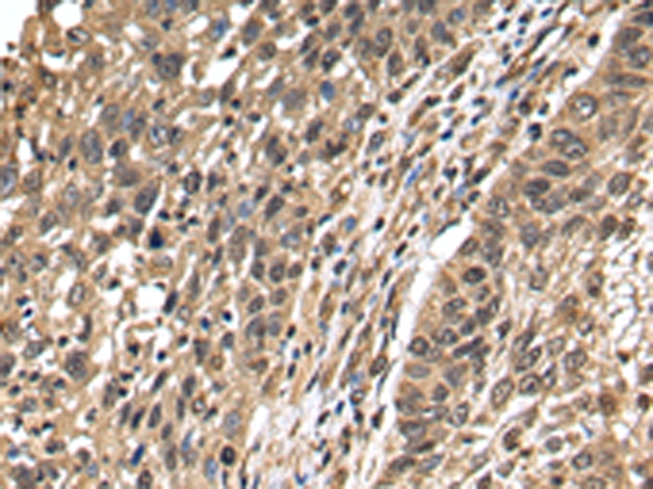
Anti-TFDP2 AntibodyA46406
ApplicationsImmunoHistoChemistry
ReactivityHuman
- SizePrice
Product group Antibodies
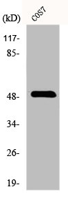
Anti-TFDP2 (Center) Antibody102-24739
ApplicationsWestern Blot
TargetTFDP2
- SizePrice

Product group Antibodies
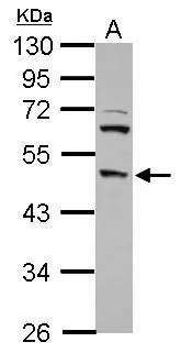
Anti-DP2/TFDP2 Antibody Picoband(r)A06850-3-CARRIER-FREE
ApplicationsWestern Blot
ReactivityMouse, Rat
TargetTFDP2
- SizePrice
Product group Antibodies
References
TFDP2 Polyclonal AntibodyBS-7660R
ApplicationsImmunoFluorescence, Western Blot, ELISA, ImmunoCytoChemistry, ImmunoHistoChemistry, ImmunoHistoChemistry Frozen, ImmunoHistoChemistry Paraffin
ReactivityBovine, Canine, Chicken, Equine, Human, Mouse, Porcine, Rabbit, Rat, Sheep
TargetTFDP2
- SizePrice
Product group Antibodies
TFDP2 AntibodyCSB-PA002187
ApplicationsWestern Blot, ELISA
ReactivityHuman, Monkey, Mouse, Rat
TargetTFDP2
- SizePrice
Product group Antibodies
Tfdp2 Polyclonal AntibodyCAC07733
ApplicationsWestern Blot, ELISA, ImmunoHistoChemistry
ReactivityMouse
TargetTFDP2
- SizePrice
Product group Antibodies
DP2 antibody [N2C1], InternalGTX102365
ApplicationsWestern Blot
ReactivityHuman, Mouse
TargetTFDP2
- SizePrice
Product group Antibodies

TFDP2 / DP2 Antibody (Biotin)LS-C500923
ApplicationsELISA
ReactivityHuman
TargetTFDP2
- SizePrice