


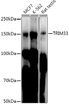
Figure 1. Western blot analysis of TIF1 gamma using anti-TIF1 gamma antibody (PB9836). Electrophoresis was performed on a 5-20% SDS-PAGE gel at 70V (Stacking gel) / 90V (Resolving gel) for 2-3 hours. The sample well of each lane was loaded with 30 ug of sample under reducing conditions. Lane 1: human K562 whole cell lysates, Lane 2: human Jurkat whole cell lysates, Lane 3: rat brain tissue lysates, Lane 4: mouse brain tissue lysates. After electrophoresis, proteins were transferred to a nitrocellulose membrane at 150 mA for 50-90 minutes. Blocked the membrane with 5% non-fat milk/TBS for 1.5 hour at RT. The membrane was incubated with rabbit anti-TIF1 gamma antigen affinity purified polyclonal antibody (Catalog # PB9836) at 0.5 microg/mL overnight at 4°C, then washed with TBS-0.1%Tween 3 times with 5 minutes each and probed with a goat anti-rabbit IgG-HRP secondary antibody at a dilution of 1:5000 for 1.5 hour at RT. The signal is developed using an Enhanced Chemiluminescent detection (ECL) kit (Catalog # EK1002) with Tanon 5200 system. A specific band was detected for TIF1 gamma at approximately 150 kDa. The expected band size for TIF1 gamma is at 123 kDa.
. TIF1 gamma was detected in a paraffin-embedded section of human lung adenocarcinoma tissue. Heat mediated antigen retrieval was performed in EDTA buffer (pH 8.0, epitope retrieval solution). The tissue section was blocked with 10% goat serum. The tissue section was then incubated with 2 microg/ml rabbit anti-TIF1 gamma Antibody (PB9836) overnight at 4°C. Peroxidase Conjugated Goat Anti-rabbit IgG was used as secondary antibody and incubated for 30 minutes at 37°C. The tissue section was developed using HRP Conjugated Rabbit IgG Super Vision Assay Kit (Catalog # SV0002) with DAB as the chromogen.")
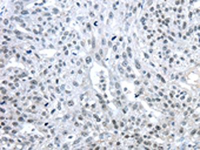
. TIF1 gamma was detected in a paraffin-embedded section of human ovarian serous adenocarcinoma tissue. Heat mediated antigen retrieval was performed in EDTA buffer (pH 8.0, epitope retrieval solution). The tissue section was blocked with 10% goat serum. The tissue section was then incubated with 2 microg/ml rabbit anti-TIF1 gamma Antibody (PB9836) overnight at 4°C. Peroxidase Conjugated Goat Anti-rabbit IgG was used as secondary antibody and incubated for 30 minutes at 37°C. The tissue section was developed using HRP Conjugated Rabbit IgG Super Vision Assay Kit (Catalog # SV0002) with DAB as the chromogen.")
. TIF1 gamma was detected in immunocytochemical section of A431 cells. Enzyme antigen retrieval was performed using IHC enzyme antigen retrieval reagent (AR0022) for 15 mins. The cells were blocked with 10% goat serum. And then incubated with 2microg/mL rabbit anti-TIF1 gamma Antibody (PB9836) overnight at 4°C. DyLight®488 Conjugated Goat Anti-Rabbit IgG (BA1127) was used as secondary antibody at 1:100 dilution and incubated for 30 minutes at 37°C. The section was counterstained with DAPI. Visualize using a fluorescence microscope and filter sets appropriate for the label used.")
. Overlay histogram showing U87 cells stained with PB9836 (Blue line). To facilitate intracellular staining, cells were fixed with 4% paraformaldehyde and permeabilized with permeabilization buffer. The cells were blocked with 10% normal goat serum. And then incubated with rabbit anti-TIF1 gamma Antibody (PB9836,1microg/1x106 cells) for 30 min at 20°C. DyLight®488 conjugated goat anti-rabbit IgG (BA1127, 5-10microg/1x106 cells) was used as secondary antibody for 30 minutes at 20°C. Isotype control antibody (Green line) was rabbit IgG (1microg/1x106) used under the same conditions. Unlabelled sample without incubation with primary antibody and secondary antibody (Red line) was used as a blank control.")
. Overlay histogram showing A431 cells stained with PB9836 (Blue line). To facilitate intracellular staining, cells were fixed with 4% paraformaldehyde and permeabilized with permeabilization buffer. The cells were blocked with 10% normal goat serum. And then incubated with rabbit anti-TIF1 gamma Antibody (PB9836,1microg/1x106 cells) for 30 min at 20°C. DyLight®488 conjugated goat anti-rabbit IgG (BA1127, 5-10microg/1x106 cells) was used as secondary antibody for 30 minutes at 20°C. Isotype control antibody (Green line) was rabbit IgG (1microg/1x106) used under the same conditions. Unlabelled sample without incubation with primary antibody and secondary antibody (Red line) was used as a blank control.")
Figure 1. Western blot analysis of TIF1 gamma using anti-TIF1 gamma antibody (PB9836). Electrophoresis was performed on a 5-20% SDS-PAGE gel at 70V (Stacking gel) / 90V (Resolving gel) for 2-3 hours. The sample well of each lane was loaded with 30 ug of sample under reducing conditions. Lane 1: human K562 whole cell lysates, Lane 2: human Jurkat whole cell lysates, Lane 3: rat brain tissue lysates, Lane 4: mouse brain tissue lysates. After electrophoresis, proteins were transferred to a nitrocellulose membrane at 150 mA for 50-90 minutes. Blocked the membrane with 5% non-fat milk/TBS for 1.5 hour at RT. The membrane was incubated with rabbit anti-TIF1 gamma antigen affinity purified polyclonal antibody (Catalog # PB9836) at 0.5 microg/mL overnight at 4°C, then washed with TBS-0.1%Tween 3 times with 5 minutes each and probed with a goat anti-rabbit IgG-HRP secondary antibody at a dilution of 1:5000 for 1.5 hour at RT. The signal is developed using an Enhanced Chemiluminescent detection (ECL) kit (Catalog # EK1002) with Tanon 5200 system. A specific band was detected for TIF1 gamma at approximately 150 kDa. The expected band size for TIF1 gamma is at 123 kDa.
Anti-TIF1 gamma/TRIM33 Antibody Picoband(r)
PB9836-CARRIER-FREE
ApplicationsFlow Cytometry, ImmunoFluorescence, Western Blot, ImmunoCytoChemistry, ImmunoHistoChemistry
Product group Antibodies
ReactivityBovine, Canine, Equine, Hamster, Human, Monkey, Mouse, Rabbit, Rat
TargetTRIM33
Overview
- SupplierBoster Bio
- Product NameAnti-TIF1 gamma/TRIM33 Antibody Picoband(r)
- Delivery Days Customer9
- Application Supplier NoteTested Species: In-house tested species with positive results. By Heat: Boiling the paraffin sections in 10mM citrate buffer, pH6.0, for 20mins is required for the staining of formalin/paraffin sections. Other applications have not been tested. Optimal dilutions should be determined by end users.
- ApplicationsFlow Cytometry, ImmunoFluorescence, Western Blot, ImmunoCytoChemistry, ImmunoHistoChemistry
- CertificationResearch Use Only
- ClonalityPolyclonal
- Concentration500 ug/ml
- Gene ID51592
- Target nameTRIM33
- Target descriptiontripartite motif containing 33
- Target synonymsDDH4, ECTO, PTC7, RFG7, TF1G, TIF1G, TIF1GAMMA, TIFGAMMA, E3 ubiquitin-protein ligase TRIM33, RET-fused gene 7 protein, RING-type E3 ubiquitin transferase TRIM33, TIF1-gamma, ectodermin homolog, protein Rfg7, transcriptional intermediary factor 1 gamma
- HostRabbit
- IsotypeIgG
- Protein IDQ9UPN9
- Protein NameE3 ubiquitin-protein ligase TRIM33
- Scientific DescriptionBoster Bio Anti-TIF1 gamma/TRIM33 Antibody Picoband® catalog # PB9836. Tested in Flow Cytometry, IF, IHC, ICC, WB applications. This antibody reacts with Human, Mouse, Rat. The brand Picoband indicates this is a premium antibody that guarantees superior quality, high affinity, and strong signals with minimal background in Western blot applications. Only our best-performing antibodies are designated as Picoband, ensuring unmatched performance.
- ReactivityBovine, Canine, Equine, Hamster, Human, Monkey, Mouse, Rabbit, Rat
- Storage Instruction-20°C,2°C to 8°C
- UNSPSC12352203
Related products
Product group Antibodies
Anti-TRIM33 AntibodyA306414
ApplicationsWestern Blot
ReactivityHuman, Rat
- SizePrice
Product group Antibodies
Anti-TRIM33 (C-term) Antibody102-21552
ApplicationsWestern Blot
TargetTRIM33
- SizePrice
Product group Antibodies

TRIM33 / TIF1-Gamma AntibodyLS-C771416
ApplicationsELISA, ImmunoHistoChemistry
ReactivityHuman, Mouse
TargetTRIM33
- SizePrice
Product group Antibodies
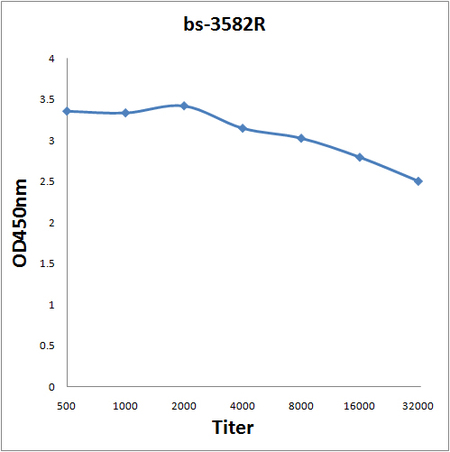
TIF1 gamma Polyclonal AntibodyBS-3582R
ApplicationsFlow Cytometry, ImmunoFluorescence, ELISA, ImmunoCytoChemistry, ImmunoHistoChemistry, ImmunoHistoChemistry Frozen, ImmunoHistoChemistry Paraffin
ReactivityBovine, Canine, Chicken, Equine, Human, Mouse, Porcine, Rat
TargetTRIM33
- SizePrice
Product group Antibodies
TRIM33 AntibodyCSB-PA104649
ApplicationsELISA, ImmunoHistoChemistry
ReactivityHuman, Mouse
TargetTRIM33
- SizePrice
Product group Antibodies
TRIM33 Polyclonal AntibodyCAC13544
ApplicationsImmunoFluorescence, ELISA, ImmunoHistoChemistry
TargetTRIM33
- SizePrice

Product group Antibodies
Anti-TRIM33 AntibodyHPA004345
ApplicationsWestern Blot, ImmunoCytoChemistry, ImmunoHistoChemistry
ReactivityHuman, Mouse, Rat
TargetTRIM33
- SizePrice
![TIF1 gamma antibody detects TIF1 gamma protein at nucleus by immunofluorescent analysis. Sample: MCF-7 cells were fixed in 4% paraformaldehyde at RT for 15 min. Green: TIF1 gamma stained by TIF1 gamma antibody (GTX131686) diluted at 1:2000. Red: alpha Tubulin, a cytoskeleton marker, stained by alpha Tubulin antibody [GT114] (GTX628802) diluted at 1:500.](https://www.genetex.com/upload/website/prouct_img/normal/GTX131686/GTX131686_42984_20180523_ICC_IF_w_23060523_526.webp)
Product group Antibodies
TIF1 gamma antibodyGTX131686
ApplicationsImmunoFluorescence, Western Blot, ImmunoCytoChemistry, ImmunoHistoChemistry, ImmunoHistoChemistry Paraffin
ReactivityHuman
TargetTRIM33
- SizePrice