


Immunohistochemical staining of human cerebral cortex shows moderate positivity in glial cells.
Immunohistochemical staining of human cerebral cortex shows moderate positivity in glial cells.
Anti-TMEM132B Antibody
HPA035661
ApplicationsImmunoHistoChemistry
Product group Antibodies
ReactivityHuman
TargetTMEM132B
Overview
- SupplierAtlas Antibodies
- Product NameAnti-TMEM132B Antibody
- Delivery Days Customer4
- ApplicationsImmunoHistoChemistry
- CertificationResearch Use Only
- ClonalityPolyclonal
- ConjugateUnconjugated
- Gene ID114795
- Target nameTMEM132B
- Target descriptiontransmembrane protein 132B
- Target synonymstransmembrane protein 132B
- HostRabbit
- IsotypeIgG
- Protein IDQ14DG7
- Protein NameTransmembrane protein 132B
- Scientific DescriptionRecombinant Protein Epitope Signature Tag (PrEST) antigen sequence
- ReactivityHuman
- Storage Instruction-20°C,2°C to 8°C
- UNSPSC41116161
Datasheet
MSDS
Related products

Product group Antibodies
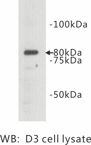
Anti-TMEM132B Antibody Picoband(r)A16132-1-CARRIER-FREE
ApplicationsFlow Cytometry, ImmunoFluorescence, Western Blot, ELISA, ImmunoHistoChemistry
ReactivityHuman, Mouse, Rat
TargetTMEM132B
- SizePrice

Product group Antibodies
Anti-TMEM132B AntibodyHPA035662
ApplicationsImmunoHistoChemistry
ReactivityHuman
TargetTMEM132B
- SizePrice
Product group Antibodies

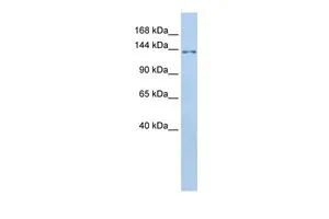
TMEM132B Antibody (aa828-877)LS-C102356
ApplicationsWestern Blot
ReactivityHuman
TargetTMEM132B
- SizePrice
Product group Antibodies
TMEM132B antibody, InternalGTX45896
ApplicationsWestern Blot
ReactivityHuman
TargetTMEM132B
- SizePrice