


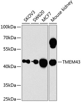
Figure 1. Western blot analysis of TMEM43 using anti-TMEM43 antibody (M05893). Electrophoresis was performed on a 5-20% SDS-PAGE gel at 70V (Stacking gel) / 90V (Resolving gel) for 2-3 hours. The sample well of each lane was loaded with 30 ug of sample under reducing conditions. Lane 1: human Jurkat whole cell lysates, Lane 2: human Hacat whole cell lysates, Lane 3: human SiHa whole cell lysates, Lane 4: human PC-3 whole cell lysates, Lane 5: rat C6 whole cell lysates, Lane 6: mouse Neuro-2a whole cell lysates. After electrophoresis, proteins were transferred to a nitrocellulose membrane at 150 mA for 50-90 minutes. Blocked the membrane with 5% non-fat milk/TBS for 1.5 hour at RT. The membrane was incubated with rabbit anti-TMEM43 antigen affinity purified monoclonal antibody (Catalog # M05893) at 1:500 overnight at 4°C, then washed with TBS-0.1%Tween 3 times with 5 minutes each and probed with a goat anti-rabbit IgG-HRP secondary antibody at a dilution of 1:1000 for 1.5 hour at RT. The signal is developed using an Enhanced Chemiluminescent detection (ECL) kit (Catalog # EK1002) with Tanon 5200 system. A specific band was detected for TMEM43 at approximately 45 kDa. The expected band size for TMEM43 is at 45 kDa.
Figure 1. Western blot analysis of TMEM43 using anti-TMEM43 antibody (M05893). Electrophoresis was performed on a 5-20% SDS-PAGE gel at 70V (Stacking gel) / 90V (Resolving gel) for 2-3 hours. The sample well of each lane was loaded with 30 ug of sample under reducing conditions. Lane 1: human Jurkat whole cell lysates, Lane 2: human Hacat whole cell lysates, Lane 3: human SiHa whole cell lysates, Lane 4: human PC-3 whole cell lysates, Lane 5: rat C6 whole cell lysates, Lane 6: mouse Neuro-2a whole cell lysates. After electrophoresis, proteins were transferred to a nitrocellulose membrane at 150 mA for 50-90 minutes. Blocked the membrane with 5% non-fat milk/TBS for 1.5 hour at RT. The membrane was incubated with rabbit anti-TMEM43 antigen affinity purified monoclonal antibody (Catalog # M05893) at 1:500 overnight at 4°C, then washed with TBS-0.1%Tween 3 times with 5 minutes each and probed with a goat anti-rabbit IgG-HRP secondary antibody at a dilution of 1:1000 for 1.5 hour at RT. The signal is developed using an Enhanced Chemiluminescent detection (ECL) kit (Catalog # EK1002) with Tanon 5200 system. A specific band was detected for TMEM43 at approximately 45 kDa. The expected band size for TMEM43 is at 45 kDa.
Anti-TMEM43 Rabbit Monoclonal Antibody
M05893
ApplicationsWestern Blot, ImmunoHistoChemistry
Product group Antibodies
ReactivityHuman, Mouse, Rat
TargetTMEM43
Overview
- SupplierBoster Bio
- Product NameAnti-TMEM43 Rabbit Monoclonal Antibody
- Delivery Days Customer9
- ApplicationsWestern Blot, ImmunoHistoChemistry
- CertificationResearch Use Only
- ClonalityMonoclonal
- Clone ID30T30
- Gene ID79188
- Target nameTMEM43
- Target descriptiontransmembrane protein 43
- Target synonymsARVC5, ARVD5, AUNA3, EDMD7, EDMD7; AUNA2, LUMA, transmembrane protein 43
- HostRabbit
- IsotypeIgG
- Protein IDQ9BTV4
- Protein NameTransmembrane protein 43
- Scientific DescriptionBoster Bio Anti-TMEM43 Rabbit Monoclonal Antibody catalog # M05893. Tested in WB, IHC applications. This antibody reacts with Human, Mouse, Rat.
- ReactivityHuman, Mouse, Rat
- Storage Instruction-20°C
- UNSPSC12352203
Related products
Product group Antibodies
Anti-TMEM43 AntibodyA12271
ApplicationsWestern Blot
ReactivityHuman, Mouse
- SizePrice
Product group Antibodies
Anti-TMEM43 Antibody144-08509
ApplicationsWestern Blot, ImmunoHistoChemistry
ReactivityHuman, Mouse
TargetTMEM43
- SizePrice

Product group Antibodies
TMEM43 Recombinant AntibodyBSM-62738R
ApplicationsImmunoFluorescence, Western Blot, ImmunoHistoChemistry, ImmunoHistoChemistry Frozen, ImmunoHistoChemistry Paraffin
ReactivityHuman, Mouse, Rat
TargetTMEM43
- SizePrice
Product group Antibodies
TMEM43 Polyclonal AntibodyCAC14885
ApplicationsWestern Blot, ELISA, ImmunoHistoChemistry
ReactivityMouse
TargetTMEM43
- SizePrice
Product group Antibodies
TMEM43 AntibodyCSB-PA023840ESR1HU
ApplicationsELISA, ImmunoHistoChemistry
ReactivityHuman
TargetTMEM43
- SizePrice
Product group Antibodies

TMEM43 AntibodyLS-C410044
ApplicationsWestern Blot
ReactivityHuman, Mouse
TargetTMEM43
- SizePrice
Product group Antibodies
TMEM43 antibodyGTX64750
ApplicationsWestern Blot, ImmunoHistoChemistry, ImmunoHistoChemistry Paraffin
ReactivityHuman, Mouse
TargetTMEM43
- SizePrice
Product group Antibodies
TargetTMEM43
- SizePrice

Product group Antibodies
Anti-TMEM43 AntibodyHPA019198
ApplicationsImmunoHistoChemistry
ReactivityHuman
TargetTMEM43
- SizePrice