

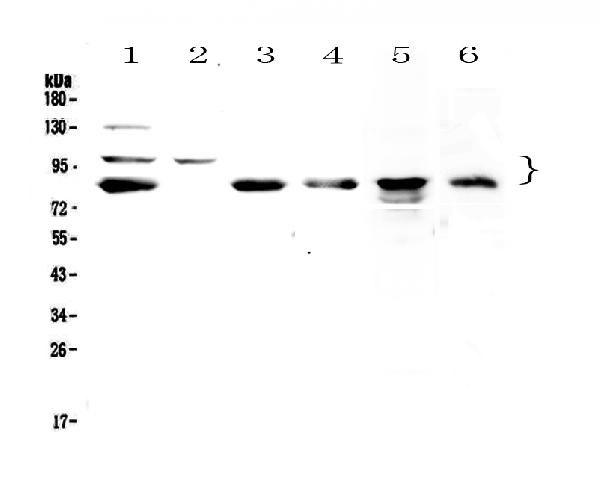
Figure 1. Western blot analysis of TPX2 using anti-TPX2 antibody (A01610-1). Electrophoresis was performed on a 5-20% SDS-PAGE gel at 70V (Stacking gel) / 90V (Resolving gel) for 2-3 hours. The sample well of each lane was loaded with 50ug of sample under reducing conditions. Lane 1: human Hela whole cell lysates, Lane 2: human COLO-320 whole cell lysates, Lane 3: human U-87MG whole cell lysates, Lane 4: human SGC-7901 whole cell lysates, Lane 5: rat PC-12 whole cell lysates, Lane 6: mouse HEPA1-6 whole cell lysates. After Electrophoresis, proteins were transferred to a Nitrocellulose membrane at 150mA for 50-90 minutes. Blocked the membrane with 5% Non-fat Milk/ TBS for 1.5 hour at RT. The membrane was incubated with rabbit anti-TPX2 antigen affinity purified polyclonal antibody (Catalog # A01610-1) at 0.5 microg/mL overnight at 4°C, then washed with TBS-0.1%Tween 3 times with 5 minutes each and probed with a goat anti-rabbit IgG-HRP secondary antibody at a dilution of 1:10000 for 1.5 hour at RT. The signal is developed using an Enhanced Chemiluminescent detection (ECL) kit (Catalog # EK1002) with Tanon 5200 system. A specific band was detected for TPX2 at approximately 86KD, 100KD. The expected band size for TPX2 is at 86KD.
Figure 1. Western blot analysis of TPX2 using anti-TPX2 antibody (A01610-1). Electrophoresis was performed on a 5-20% SDS-PAGE gel at 70V (Stacking gel) / 90V (Resolving gel) for 2-3 hours. The sample well of each lane was loaded with 50ug of sample under reducing conditions. Lane 1: human Hela whole cell lysates, Lane 2: human COLO-320 whole cell lysates, Lane 3: human U-87MG whole cell lysates, Lane 4: human SGC-7901 whole cell lysates, Lane 5: rat PC-12 whole cell lysates, Lane 6: mouse HEPA1-6 whole cell lysates. After Electrophoresis, proteins were transferred to a Nitrocellulose membrane at 150mA for 50-90 minutes. Blocked the membrane with 5% Non-fat Milk/ TBS for 1.5 hour at RT. The membrane was incubated with rabbit anti-TPX2 antigen affinity purified polyclonal antibody (Catalog # A01610-1) at 0.5 microg/mL overnight at 4°C, then washed with TBS-0.1%Tween 3 times with 5 minutes each and probed with a goat anti-rabbit IgG-HRP secondary antibody at a dilution of 1:10000 for 1.5 hour at RT. The signal is developed using an Enhanced Chemiluminescent detection (ECL) kit (Catalog # EK1002) with Tanon 5200 system. A specific band was detected for TPX2 at approximately 86KD, 100KD. The expected band size for TPX2 is at 86KD.
Anti-TPX2 Antibody Picoband(r)
A01610-1-APC
ApplicationsWestern Blot, ELISA
Product group Antibodies
ReactivityHuman, Mouse, Rat
TargetTPX2
Overview
- SupplierBoster Bio
- Product NameAnti-TPX2 Antibody Picoband(r)
- Delivery Days Customer9
- ApplicationsWestern Blot, ELISA
- CertificationResearch Use Only
- ClonalityPolyclonal
- Concentration500 ug/ml
- ConjugateAPC (Allophycocyanin)
- Gene ID22974
- Target nameTPX2
- Target descriptionTPX2 microtubule nucleation factor
- Target synonymsC20orf1, C20orf2, DIL-2, DIL2, FLS353, GD:C20orf1, HCA519, HCTP4, REPP86, p100, targeting protein for Xklp2, TPX2, microtubule-associated protein homolog, TPX2, microtubule-associated, homolog, differentially expressed in cancerous and non-cancerous lung cells 2, differentially expressed in cancerous and noncancerous lung cells 2, differentially expressed in lung cells, hepatocellular carcinoma-associated antigen 519, hepatocellular carcinoma-associated antigen 90, preferentially expressed in colorectal cancer, protein fls353, restricted expression proliferation associated protein 100
- HostRabbit
- IsotypeIgG
- Protein IDQ9ULW0
- Protein NameTargeting protein for Xklp2
- Scientific DescriptionBoster Bio Anti-TPX2 Antibody Picoband® catalog # A01610-1. Tested in ELISA, WB applications. This antibody reacts with Human, Mouse, Rat. The brand Picoband indicates this is a premium antibody that guarantees superior quality, high affinity, and strong signals with minimal background in Western blot applications. Only our best-performing antibodies are designated as Picoband, ensuring unmatched performance.
- ReactivityHuman, Mouse, Rat
- Storage Instruction-20°C,2°C to 8°C
- UNSPSC12352203