


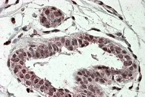
Immunohistochemical staining of human Skin shows moderate nuclear positivity in squamous epithelial cells.
Immunohistochemical staining of human Skin shows moderate nuclear positivity in squamous epithelial cells.
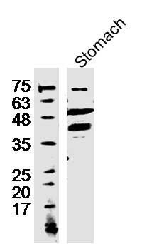
Anti-TREX2 Antibody
HPA054060
ApplicationsImmunoHistoChemistry
Product group Antibodies
ReactivityHuman
TargetTREX2
Overview
- SupplierAtlas Antibodies
- Product NameAnti-TREX2 Antibody
- Delivery Days Customer4
- ApplicationsImmunoHistoChemistry
- CertificationResearch Use Only
- ClonalityPolyclonal
- ConjugateUnconjugated
- Gene ID11219
- Target nameTREX2
- Target descriptionthree prime repair exonuclease 2
- Target synonymsthree prime repair exonuclease 2, 3'-5' exonuclease TREX2 long form
- HostRabbit
- IsotypeIgG
- Protein IDQ9BQ50
- Protein NameThree prime repair exonuclease 2
- Scientific DescriptionRecombinant Protein Epitope Signature Tag (PrEST) antigen sequence
- ReactivityHuman
- Storage Instruction-20°C,2°C to 8°C
- UNSPSC41116161
Datasheet
MSDS
Related products

Product group Antibodies
Anti-TREX2 AntibodyA47972
ApplicationsWestern Blot, ELISA, ImmunoHistoChemistry
ReactivityHuman, Mouse, Rat
- SizePrice
Product group Antibodies

Goat anti-TREX2EB08131
ApplicationsELISA, ImmunoHistoChemistry
ReactivityHuman
TargetTREX2
- SizePrice
Product group Antibodies

TREX2 AntibodyLS-C662386
ApplicationsWestern Blot
ReactivityHuman
TargetTREX2
- SizePrice

Product group Antibodies
Anti-TREX2 Antibody Picoband(r)PB10105-CARRIER-FREE
ApplicationsWestern Blot
ReactivityHuman
TargetTREX2
- SizePrice
Product group Antibodies
TREX2 antibody, InternalGTX47594
ApplicationsImmunoHistoChemistry, ImmunoHistoChemistry Paraffin
ReactivityHuman
TargetTREX2
- SizePrice
Product group Antibodies
Anti-TREX2 (C-term) Antibody102-24262
ApplicationsWestern Blot
TargetTREX2
- SizePrice
Product group Antibodies
TREX2 Polyclonal AntibodyBS-16713R
ApplicationsImmunoFluorescence, ELISA, ImmunoCytoChemistry, ImmunoHistoChemistry, ImmunoHistoChemistry Frozen, ImmunoHistoChemistry Paraffin
ReactivityBovine, Canine, Equine, Human, Mouse, Rabbit, Rat
TargetTREX2
- SizePrice