


Immunofluorescent staining of human cell line MCF7 shows localization to nucleoplasm.
Immunofluorescent staining of human cell line MCF7 shows localization to nucleoplasm.
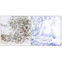
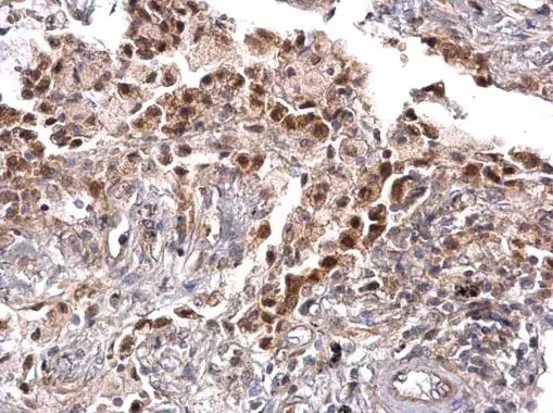
Anti-TRPS1 Antibody
HPA060380
ApplicationsImmunoCytoChemistry
Product group Antibodies
ReactivityHuman
TargetTRPS1
Overview
- SupplierAtlas Antibodies
- Product NameAnti-TRPS1 Antibody
- Delivery Days Customer4
- ApplicationsImmunoCytoChemistry
- CertificationResearch Use Only
- ClonalityPolyclonal
- ConjugateUnconjugated
- Gene ID7227
- Target nameTRPS1
- Target descriptiontranscriptional repressor GATA binding 1
- Target synonymsGC79, LGCR, zinc finger transcription factor Trps1, tricho-rhino-phalangeal syndrome type I protein, trichorhinophalangeal syndrome I, zinc finger protein GC79
- HostRabbit
- IsotypeIgG
- Protein IDQ9UHF7
- Protein NameZinc finger transcription factor Trps1
- Scientific DescriptionRecombinant Protein Epitope Signature Tag (PrEST) antigen sequence
- ReactivityHuman
- Storage Instruction-20°C,2°C to 8°C
- UNSPSC41116161
Datasheet
MSDS
Related products
Product group Antibodies
TRPS1 AntibodyCSB-PA060010
ApplicationsELISA, ImmunoHistoChemistry
ReactivityHuman, Mouse
TargetTRPS1
- SizePrice
Product group Antibodies
Anti-TRPS1 AntibodyA45821
ApplicationsImmunoHistoChemistry
ReactivityHuman, Mouse
- SizePrice

Product group Antibodies
Anti-TRPS1 Antibody Picoband(r)A02328-2-CARRIER-FREE
ApplicationsFlow Cytometry, ImmunoFluorescence, Western Blot, ELISA, ImmunoCytoChemistry, ImmunoHistoChemistry
ReactivityHuman
TargetTRPS1
- SizePrice

Product group Antibodies
Anti-TRPS1 AntibodyHPA071767
ApplicationsImmunoHistoChemistry
ReactivityHuman
TargetTRPS1
- SizePrice
Product group Antibodies

TRPS1 AntibodyLS-C409294
ApplicationsImmunoFluorescence, Western Blot
ReactivityHuman, Mouse
TargetTRPS1
- SizePrice
Product group Antibodies
TRPS1 antibodyGTX129429
ApplicationsWestern Blot, ImmunoHistoChemistry, ImmunoHistoChemistry Paraffin
ReactivityHuman
TargetTRPS1
- SizePrice
Product group Antibodies
Anti-TRPS1 Antibody144-07743
ApplicationsImmunoFluorescence, Western Blot
ReactivityHuman, Mouse
TargetTRPS1
- SizePrice