Bio-Connect
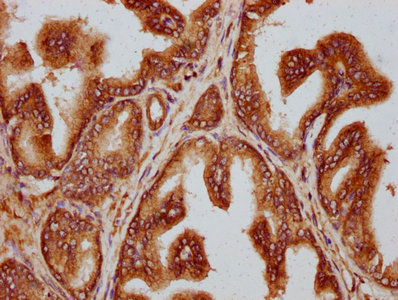
Immunohistochemical staining of human ovary shows cytoplasmic positivity in follicle cells.

Anti-TTLL3 Antibody

HPA051413
Atlas Antibodies
ApplicationsImmunoHistoChemistry
Product group Antibodies
TargetTTLL3
Sign in to order and to see your custom pricing.
Large volume orders?
Order with a bulk request

Overview

  • Supplier
    Atlas Antibodies
  • Product Name
    Anti-TTLL3
  • Delivery Days Customer
    4
  • Applications
    ImmunoHistoChemistry
  • Certification
    Research Use Only
  • Clonality
    Polyclonal
  • Conjugate
    Unconjugated
  • Gene ID26140
  • Target name
    TTLL3
  • Target description
    tubulin tyrosine ligase like 3
  • Target synonyms
    HOTTL; tubulin monoglycylase TTLL3; tubulin tyrosine ligase-like family, member 3; tubulin--tyrosine ligase-like protein 3
  • Host
    Rabbit
  • Isotype
    IgG
  • Scientific Description
    Recombinant Protein Epitope Signature Tag (PrEST) antigen sequence
  • Storage Instruction
    -20°C,2°C to 8°C
  • UNSPSC
    12352203

Related products

Atlas Antibodies
  • SizePrice
Figure 1. Western blot analysis of TTLL3 using anti-TTLL3 antibody (A11845-1). Electrophoresis was performed on a 5-20% SDS-PAGE gel at 70V (Stacking gel) / 90V (Resolving gel) for 2-3 hours. The sample well of each lane was loaded with 30 ug of sample under reducing conditions. Lane 1: human 293T whole cell lysates, Lane 2: human Hela whole cell lysates, Lane 3: human Jurkat whole cell lysates. After electrophoresis, proteins were transferred to a nitrocellulose membrane at 150 mA for 50-90 minutes. Blocked the membrane with 5% non-fat milk/TBS for 1.5 hour at RT. The membrane was incubated with rabbit anti-TTLL3 antigen affinity purified polyclonal antibody (Catalog # A11845-1) at 0.5 microg/mL overnight at 4°C, then washed with TBS-0.1%Tween 3 times with 5 minutes each and probed with a goat anti-rabbit IgG-HRP secondary antibody at a dilution of 1:5000 for 1.5 hour at RT. The signal is developed using an Enhanced Chemiluminescent detection (ECL) kit (Catalog # EK1002) with Tanon 5200 system. A specific band was detected for TTLL3 at approximately 87 kDa. The expected band size for TTLL3 is at 87 kDa.
Product group Antibodies
Boster Bio
Anti-TTLL3 Antibody Picoband(r)A11845-1-CARRIER-FREE
ApplicationsWestern Blot, ELISA
TargetTTLL3
  • SizePrice
Product group Antibodies
LifeSpan BioSciences
TTLL3 AntibodyLS-C681637
ApplicationsELISA, ImmunoHistoChemistry, ImmunoHistoChemistry Paraffin
TargetTTLL3
  • SizePrice
Product group Antibodies
Assay Genie
ApplicationsELISA, ImmunoHistoChemistry
TargetTTLL3
  • SizePrice
IHC image of CSB-PA23329A0Rb diluted at 1:300 and staining in paraffin-embedded human prostate cancer performed on a Leica BondTM system. After dewaxing and hydration, antigen retrieval was mediated by high pressure in a citrate buffer (pH 6.0). Section was blocked with 10% normal goat serum 30min at RT. Then primary antibody (1% BSA) was incubated at 4°C overnight. The primary is detected by a biotinylated secondary antibody and visualized using an HRP conjugated SP system.
Product group Antibodies
Cusabio
TTLL3 AntibodyCSB-PA23329A0RB
ApplicationsELISA, ImmunoHistoChemistry
ReactivityHuman
TargetTTLL3
  • SizePrice