


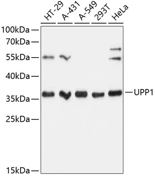
Figure 1. Western blot analysis of UPP1 using anti-UPP1 antibody (M10128). Electrophoresis was performed on a 5-20% SDS-PAGE gel at 70V (Stacking gel) / 90V (Resolving gel) for 2-3 hours. The sample well of each lane was loaded with 30 ug of sample under reducing conditions. Lane 1: human Hela whole cell lysates, Lane 2: human 293T whole cell lysates, Lane 3: human Jurkat whole cell lysates, Lane 4: human MCF-7 whole cell lysates, Lane 5: rat heart tissue lysates, Lane 6: rat liver tissue lysates, Lane 7: mouse heart tissue lysates, Lane 8: mouse liver tissue lysates. After electrophoresis, proteins were transferred to a nitrocellulose membrane at 150 mA for 50-90 minutes. Blocked the membrane with 5% non-fat milk/TBS for 1.5 hour at RT. The membrane was incubated with rabbit anti-UPP1 antigen affinity purified monoclonal antibody (M10128) at 1:500 overnight at 4°C, then washed with TBS-0.1%Tween 3 times with 5 minutes each and probed with a goat anti-rabbit IgG-HRP secondary antibody at a dilution of 1:500 for 1.5 hour at RT. The signal is developed using an Enhanced Chemiluminescent detection (ECL) kit (Catalog # EK1002) with Tanon 5200 system. A specific band was detected for UPP1 at approximately 28 kDa. The expected band size for UPP1 is at 34 kDa.
Figure 1. Western blot analysis of UPP1 using anti-UPP1 antibody (M10128). Electrophoresis was performed on a 5-20% SDS-PAGE gel at 70V (Stacking gel) / 90V (Resolving gel) for 2-3 hours. The sample well of each lane was loaded with 30 ug of sample under reducing conditions. Lane 1: human Hela whole cell lysates, Lane 2: human 293T whole cell lysates, Lane 3: human Jurkat whole cell lysates, Lane 4: human MCF-7 whole cell lysates, Lane 5: rat heart tissue lysates, Lane 6: rat liver tissue lysates, Lane 7: mouse heart tissue lysates, Lane 8: mouse liver tissue lysates. After electrophoresis, proteins were transferred to a nitrocellulose membrane at 150 mA for 50-90 minutes. Blocked the membrane with 5% non-fat milk/TBS for 1.5 hour at RT. The membrane was incubated with rabbit anti-UPP1 antigen affinity purified monoclonal antibody (M10128) at 1:500 overnight at 4°C, then washed with TBS-0.1%Tween 3 times with 5 minutes each and probed with a goat anti-rabbit IgG-HRP secondary antibody at a dilution of 1:500 for 1.5 hour at RT. The signal is developed using an Enhanced Chemiluminescent detection (ECL) kit (Catalog # EK1002) with Tanon 5200 system. A specific band was detected for UPP1 at approximately 28 kDa. The expected band size for UPP1 is at 34 kDa.
Anti-UPP1 Rabbit Monoclonal Antibody
M10128
ApplicationsWestern Blot
Product group Antibodies
ReactivityHuman, Mouse, Rat
TargetUPP1
Overview
- SupplierBoster Bio
- Product NameAnti-UPP1 Rabbit Monoclonal Antibody
- Delivery Days Customer9
- ApplicationsWestern Blot
- CertificationResearch Use Only
- ClonalityMonoclonal
- Clone ID26U88
- Gene ID7378
- Target nameUPP1
- Target descriptionuridine phosphorylase 1
- Target synonymsUDRPASE, UP, UPASE, UPP, uridine phosphorylase 1, UPase 1, urdPase 1
- HostRabbit
- IsotypeIgG
- Protein IDQ16831
- Protein NameUridine phosphorylase 1
- Scientific DescriptionBoster Bio Anti-UPP1 Rabbit Monoclonal Antibody catalog # M10128. Tested in WB application. This antibody reacts with Human, Mouse, Rat.
- ReactivityHuman, Mouse, Rat
- Storage Instruction-20°C
- UNSPSC12352203
Related products
Product group Antibodies

UP / UPP1 AntibodyLS-C748115
ApplicationsWestern Blot
ReactivityHuman
TargetUPP1
- SizePrice

Product group Antibodies
Anti-UPP1 AntibodyHPA054040
ApplicationsWestern Blot, ImmunoCytoChemistry
ReactivityHuman
TargetUPP1
- SizePrice
Product group Antibodies
ApplicationsImmunoPrecipitation, Western Blot, ImmunoCytoChemistry, ImmunoHistoChemistry
ReactivityMouse, Porcine
TargetUPP1
- SizePrice
Product group Antibodies
UPP1 antibodyGTX66155
ApplicationsWestern Blot
ReactivityHuman
TargetUPP1
- SizePrice
Product group Antibodies
ApplicationsWestern Blot
ReactivityHuman, Mouse, Rat
TargetUPP1
- SizePrice
Product group Antibodies
Anti-UPP1 Antibody144-60163
ApplicationsWestern Blot
ReactivityHuman
TargetUPP1
- SizePrice