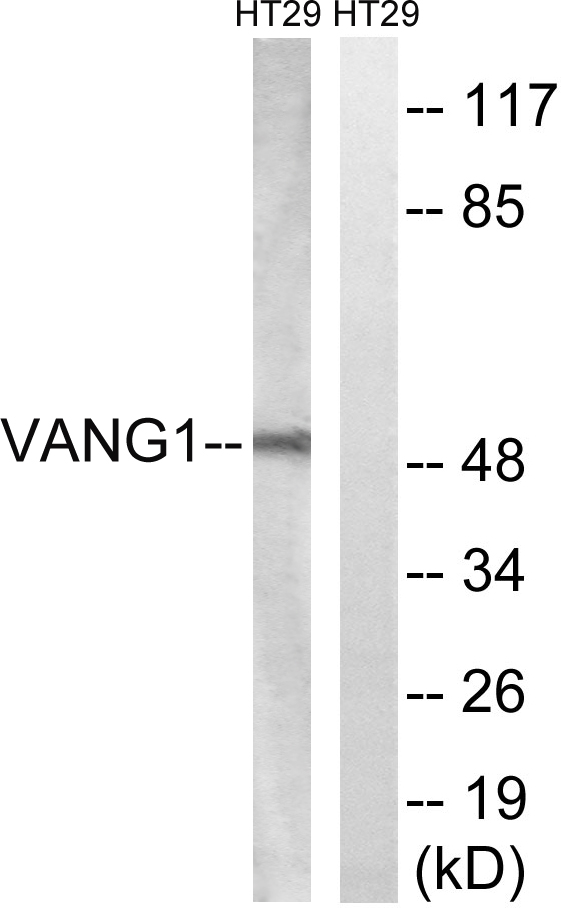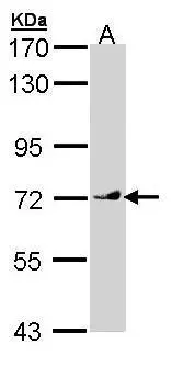
Immunofluorescent staining of MCF7 cells shows localization to plasma membrane.
Anti-VANGL1 Antibody
HPA025235
ApplicationsWestern Blot, ImmunoCytoChemistry
Product group Antibodies
ReactivityHuman
TargetVANGL1
Overview
- SupplierAtlas Antibodies
- Product NameAnti-VANGL1 Antibody
- Delivery Days Customer4
- ApplicationsWestern Blot, ImmunoCytoChemistry
- CertificationResearch Use Only
- ClonalityPolyclonal
- ConjugateUnconjugated
- Gene ID81839
- Target nameVANGL1
- Target descriptionVANGL planar cell polarity protein 1
- Target synonymsKITENIN, LPP2, STB2, STBM2, vang-like protein 1, KAI1 C-terminal interacting tetraspanin, loop-tail protein 2 homolog, strabismus 2, van Gogh-like protein 1, vang-like 1 (van gogh, Drosophila)
- HostRabbit
- IsotypeIgG
- Protein IDQ8TAA9
- Protein NameVang-like protein 1
- Scientific DescriptionRecombinant Protein Epitope Signature Tag (PrEST) antigen sequence
- ReactivityHuman
- Storage Instruction-20°C,2°C to 8°C
- UNSPSC41116161


