


Immunohistochemical staining of human placenta shows strong cytoplasmic positivity in trophoblastic cells.
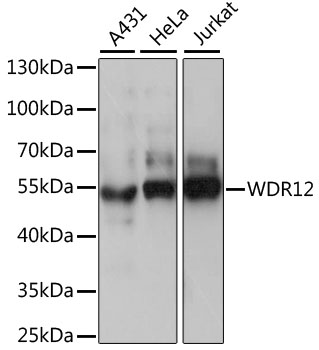
. Lane 2: NBT-II cell lysate (Rat Wistar bladder tumour cells)")
Immunohistochemical staining of human placenta shows strong cytoplasmic positivity in trophoblastic cells.
Anti-WDR12 Antibody
HPA036389
ApplicationsWestern Blot, ImmunoHistoChemistry
Product group Antibodies
ReactivityHuman, Mouse
TargetWDR12
Overview
- SupplierAtlas Antibodies
- Product NameAnti-WDR12 Antibody
- Delivery Days Customer4
- ApplicationsWestern Blot, ImmunoHistoChemistry
- CertificationResearch Use Only
- ClonalityPolyclonal
- ConjugateUnconjugated
- Gene ID55759
- Target nameWDR12
- Target descriptionWD repeat domain 12
- Target synonymsYTM1, ribosome biogenesis protein WDR12, WD repeat-containing protein 12
- HostRabbit
- IsotypeIgG
- Protein IDQ9GZL7
- Protein NameRibosome biogenesis protein WDR12
- Scientific DescriptionRecombinant Protein Epitope Signature Tag (PrEST) antigen sequence
- ReactivityHuman, Mouse
- Storage Instruction-20°C,2°C to 8°C
- UNSPSC41116161
Datasheet
MSDS
Related products
Product group Antibodies

Anti-WDR12 [RAB-C527]Ab01921-1.1
ApplicationsFlow Cytometry, ImmunoFluorescence
ReactivityHuman
TargetWDR12
- SizePrice

Product group Antibodies
Anti-WDR12 Antibody Picoband(r)A07492-1-CARRIER-FREE
ApplicationsFlow Cytometry, Western Blot, ELISA
ReactivityHuman
TargetWDR12
- SizePrice
Product group Antibodies
Anti-WDR12 Antibody144-61074
ApplicationsWestern Blot
ReactivityHuman
TargetWDR12
- SizePrice
Product group Antibodies

WDR12 AntibodyLS-C750446
ApplicationsWestern Blot
ReactivityHuman
TargetWDR12
- SizePrice
Product group Antibodies
WDR12 AntibodyCSB-PA188811XA01DOA
ApplicationsWestern Blot, ELISA
ReactivityPlant
- SizePrice
Product group Antibodies
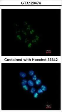
WDR12 antibody [N1C2]GTX120474
ApplicationsImmunoFluorescence, Western Blot, ImmunoCytoChemistry
ReactivityHuman, Mouse
TargetWDR12
- SizePrice
Product group Antibodies
Anti-WDR12 AntibodyCAB15477
ApplicationsWestern Blot, ELISA
ReactivityHuman
TargetWDR12
- SizePrice