


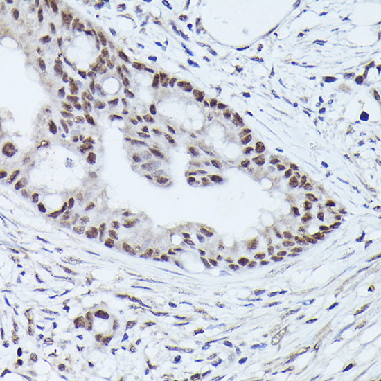
Immunohistochemical staining of human thyroid gland shows strong cytoplasmic positivity in glandular cells.
Immunohistochemical staining of human thyroid gland shows strong cytoplasmic positivity in glandular cells.
Anti-WDR46 Antibody
HPA043004
ApplicationsImmunoHistoChemistry
Product group Antibodies
ReactivityHuman
TargetWDR46
Overview
- SupplierAtlas Antibodies
- Product NameAnti-WDR46 Antibody
- Delivery Days Customer4
- ApplicationsImmunoHistoChemistry
- CertificationResearch Use Only
- ClonalityPolyclonal
- ConjugateUnconjugated
- Gene ID9277
- Target nameWDR46
- Target descriptionWD repeat domain 46
- Target synonymsBING4, C6orf11, FP221, UTP7, WD repeat-containing protein 46, WD repeat-containing protein BING4
- HostRabbit
- IsotypeIgG
- Protein IDO15213
- Protein NameWD repeat-containing protein 46
- Scientific DescriptionRecombinant Protein Epitope Signature Tag (PrEST) antigen sequence
- ReactivityHuman
- Storage Instruction-20°C,2°C to 8°C
- UNSPSC41116161
Datasheet
MSDS
Related products
Product group Antibodies
Anti-WDR46 AntibodyA305665
ApplicationsImmunoFluorescence, Western Blot, ImmunoCytoChemistry, ImmunoHistoChemistry
ReactivityHuman, Mouse, Rat
- SizePrice
Product group Antibodies
WDR46 AntibodyCSB-PA026034GA01HU
ApplicationsWestern Blot, ELISA
ReactivityHuman, Mouse, Rat
TargetWDR46
- SizePrice
Product group Antibodies

WDR46 AntibodyLS-C661606
ApplicationsWestern Blot, ELISA
ReactivityHuman
TargetWDR46
- SizePrice

Product group Antibodies
Anti-WDR46 Antibody Picoband(r)A12816-1-CARRIER-FREE
ApplicationsFlow Cytometry, Western Blot, ELISA
ReactivityHuman
TargetWDR46
- SizePrice