


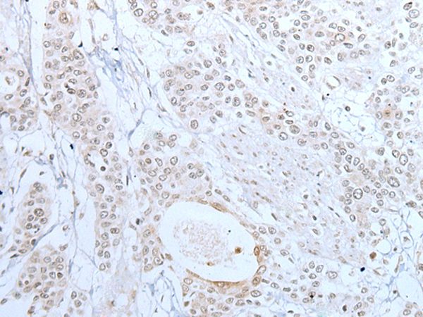
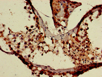
Immunohistochemical staining of human kidney shows strong cytoplasmic positivity in cells in tubules.

Immunohistochemical staining of human kidney shows strong cytoplasmic positivity in cells in tubules.
Anti-XAF1 Antibody
HPA057302
ApplicationsImmunoCytoChemistry, ImmunoHistoChemistry
Product group Antibodies
ReactivityHuman
TargetXAF1
Overview
- SupplierAtlas Antibodies
- Product NameAnti-XAF1 Antibody
- Delivery Days Customer4
- ApplicationsImmunoCytoChemistry, ImmunoHistoChemistry
- CertificationResearch Use Only
- ClonalityPolyclonal
- ConjugateUnconjugated
- Gene ID54739
- Target nameXAF1
- Target descriptionXIAP associated factor 1
- Target synonymsBIRC4BP, HSXIAPAF1, XIAPAF1, XIAP-associated factor 1, BIRC4-binding protein
- HostRabbit
- IsotypeIgG
- Protein IDQ6GPH4
- Protein NameXIAP-associated factor 1
- Scientific DescriptionRecombinant Protein Epitope Signature Tag (PrEST) antigen sequence
- ReactivityHuman
- Storage Instruction-20°C,2°C to 8°C
- UNSPSC41116161
Datasheet
MSDS
Related products
Product group Antibodies
Anti-XAF1 AntibodyA47117
ApplicationsImmunoHistoChemistry
ReactivityHuman
- SizePrice
Product group Antibodies
Anti-XAF1 (C-term) Antibody102-23874
ApplicationsImmunoFluorescence, Western Blot
TargetXAF1
- SizePrice

Product group Antibodies
Anti-XAF1 Antibody Picoband(r)A03432-1-CARRIER-FREE
ApplicationsFlow Cytometry, Western Blot, ELISA, ImmunoHistoChemistry
ReactivityHuman
TargetXAF1
- SizePrice
Product group Antibodies
XAF1 AntibodyCSB-PA764111LA01HU
ApplicationsImmunoFluorescence, ELISA, ImmunoHistoChemistry
ReactivityHuman
TargetXAF1
- SizePrice
Product group Antibodies

XAF1 AntibodyLS-C501165
ApplicationsImmunoFluorescence, ELISA, ImmunoHistoChemistry
ReactivityHuman
TargetXAF1
- SizePrice