

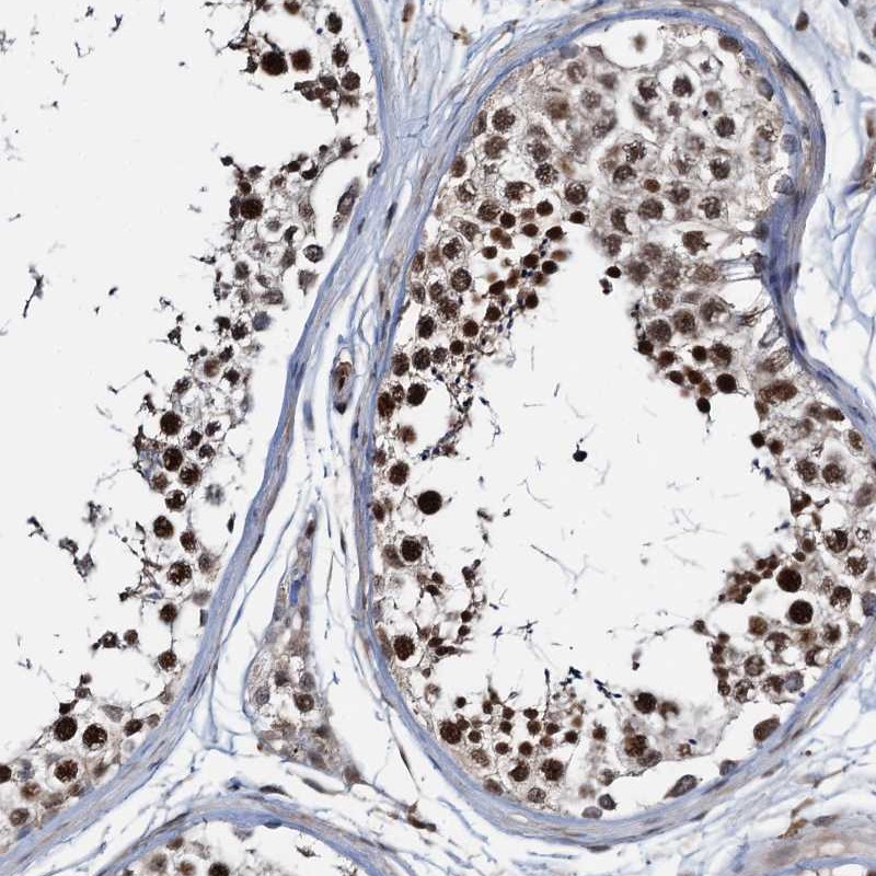
Immunohistochemical staining of human testis shows strong nuclear positivity in cells in seminiferous ducts.
![Lane 1: Marker [kDa] 250, 130, 95, 72, 55, 36, 28, 17, 10. Lane 2: Human cell line RT-4. Lane 3: Human cell line U-251MG sp](https://atlasantibodies.s3.amazonaws.com/images/wb/hpa040832-wb-1.jpg "Lane 1: Marker [kDa] 250, 130, 95, 72, 55, 36, 28, 17, 10. Lane 2: Human cell line RT-4. Lane 3: Human cell line U-251MG sp")
Immunohistochemical staining of human testis shows strong nuclear positivity in cells in seminiferous ducts.
Anti-ZNF609 Antibody
HPA040832
ApplicationsWestern Blot, ImmunoCytoChemistry, ImmunoHistoChemistry
Product group Antibodies
ReactivityHuman, Rat
TargetZNF609
Overview
- SupplierAtlas Antibodies
- Product NameAnti-ZNF609 Antibody
- Delivery Days Customer4
- ApplicationsWestern Blot, ImmunoCytoChemistry, ImmunoHistoChemistry
- CertificationResearch Use Only
- ClonalityPolyclonal
- ConjugateUnconjugated
- Gene ID23060
- Target nameZNF609
- Target descriptionzinc finger protein 609
- Target synonymszinc finger protein 609
- HostRabbit
- IsotypeIgG
- Protein IDO15014
- Protein NameZinc finger protein 609
- Scientific DescriptionRecombinant Protein Epitope Signature Tag (PrEST) antigen sequence
- ReactivityHuman, Rat
- Storage Instruction-20°C,2°C to 8°C
- UNSPSC41116161
Datasheet
MSDS
Related products

Product group Antibodies
Anti-ZNF609 Antibody Picoband(r)A15674-1-CARRIER-FREE
ApplicationsFlow Cytometry, Western Blot, ELISA
ReactivityHuman
TargetZNF609
- SizePrice
Product group Antibodies

ZNF609 Antibody (aa71-120)LS-C31487
ApplicationsWestern Blot
ReactivityHuman
TargetZNF609
- SizePrice

Product group Antibodies
Anti-ZNF609 AntibodyHPA040742
ApplicationsChIP Chromatin ImmunoPrecipitation, ImmunoCytoChemistry, ImmunoHistoChemistry
ReactivityHuman
TargetZNF609
- SizePrice