

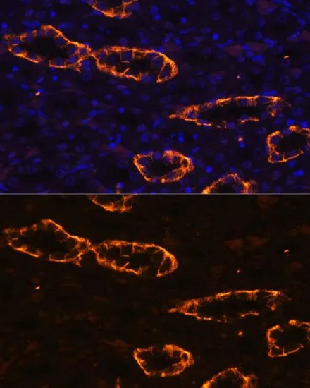
IHC-P analysis of rat kidney tissue using GTX64481 Aquaporin 3 antibody. Blue : DAPI Dilution : 1:100


IHC-P analysis of rat kidney tissue using GTX64481 Aquaporin 3 antibody. Blue : DAPI Dilution : 1:100
Aquaporin 3 antibody
GTX64481
ApplicationsWestern Blot, ImmunoHistoChemistry, ImmunoHistoChemistry Paraffin
Product group Antibodies
ReactivityHuman, Mouse, Rat
TargetAQP3
Overview
- SupplierGeneTex
- Product NameAquaporin 3 antibody
- Delivery Days Customer9
- Application Supplier NoteWB: 1:500 - 1:2000. IHC-P: 1:50 - 1:200. *Optimal dilutions/concentrations should be determined by the researcher.Not tested in other applications.
- ApplicationsWestern Blot, ImmunoHistoChemistry, ImmunoHistoChemistry Paraffin
- CertificationResearch Use Only
- ClonalityPolyclonal
- ConjugateUnconjugated
- Gene ID360
- Target nameAQP3
- Target descriptionaquaporin 3 (Gill blood group)
- Target synonymsAQP-3, GIL, aquaporin-3, aquaglyceroporin-3, aquaporin 3 (GIL blood group)
- HostRabbit
- IsotypeIgG
- Protein IDQ92482
- Protein NameAquaporin-3
- Scientific DescriptionThis gene encodes the water channel protein aquaporin 3. Aquaporins are a family of small integral membrane proteins related to the major intrinsic protein, also known as aquaporin 0. Aquaporin 3 is localized at the basal lateral membranes of collecting duct cells in the kidney. In addition to its water channel function, aquaporin 3 has been found to facilitate the transport of nonionic small solutes such as urea and glycerol, but to a smaller degree. It has been suggested that water channels can be functionally heterogeneous and possess water and solute permeation mechanisms. Alternative splicing of this gene results in multiple transcript variants encoding different isoforms. [provided by RefSeq, Dec 2015]
- ReactivityHuman, Mouse, Rat
- Storage Instruction-20°C or -80°C,2°C to 8°C
- UNSPSC41116161
Datasheet
Related products
Product group Antibodies
Anti-AQP3 AntibodyA97793
ApplicationsELISA, ImmunoHistoChemistry
ReactivityHuman, Mouse, Rat
- SizePrice
Product group Antibodies
Anti-AQP3 Antibody144-02838
ApplicationsImmunoFluorescence, Western Blot, ImmunoHistoChemistry
ReactivityHuman, Mouse, Rat
TargetAQP3
- SizePrice
Product group Antibodies

AQP3 / Aquaporin 3 AntibodyLS-C764594
ApplicationsWestern Blot, ELISA, ImmunoHistoChemistry
ReactivityHuman, Mouse, Rat
TargetAQP3
- SizePrice
Product group Antibodies
AQP3 AntibodyCSB-PA009603
ApplicationsELISA, ImmunoHistoChemistry
ReactivityHuman, Mouse, Rat
TargetAQP3
- SizePrice
Product group Antibodies
Aqp3 Polyclonal AntibodyCAC10970
ApplicationsImmunoFluorescence, ELISA, ImmunoHistoChemistry
TargetAQP3
- SizePrice

Product group Antibodies
Anti-AQP3 AntibodyHPA014924
ApplicationsImmunoCytoChemistry, ImmunoHistoChemistry
ReactivityHuman
TargetAQP3
- SizePrice

Product group Antibodies
Anti-Aquaporin 3/AQP3 Antibody Picoband(r)PA1488-CARRIER-FREE
ApplicationsFlow Cytometry, ImmunoFluorescence, Western Blot, ImmunoCytoChemistry, ImmunoHistoChemistry
ReactivityBovine, Human, Mouse, Rat
TargetAQP3
- SizePrice