

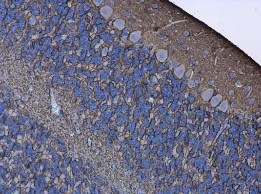
Aquaporin 4 antibody detects Aquaporin 4 protein at cell membrane in mouse brain by immunohistochemical analysis. Sample: Paraffin-embedded mouse brain. Aquaporin 4 antibody (GTX133151) diluted at 1:500.
Antigen Retrieval: Citrate buffer, pH 6.0, 15 min
![Aquaporin 4 antibody detects Aquaporin 4 protein at astrocyte cells by immunofluorescent analysis. Sample: DIV9 rat E18 primary cortical neurons and glia cells were fixed in 4% paraformaldehyde at RT for 15 min. Green: Aquaporin 4 protein stained by Aquaporin 4 antibody (GTX133151) diluted at 1:500. Red: beta Tubulin 3/ Tuj1, a neuron cell marker, stained by beta Tubulin 3/ Tuj1 antibody [GT886] (GTX631830) diluted at 1:500. Blue: Fluoroshield with DAPI (GTX30920).](https://www.genetex.com/upload/website/prouct_img/normal/GTX133151/GTX133151_42640_20170705_IFA_R_w_23060523_778.webp "Aquaporin 4 antibody detects Aquaporin 4 protein at astrocyte cells by immunofluorescent analysis. Sample: DIV9 rat E18 primary cortical neurons and glia cells were fixed in 4% paraformaldehyde at RT for 15 min. Green: Aquaporin 4 protein stained by Aquaporin 4 antibody (GTX133151) diluted at 1:500. Red: beta Tubulin 3/ Tuj1, a neuron cell marker, stained by beta Tubulin 3/ Tuj1 antibody [GT886] (GTX631830) diluted at 1:500. Blue: Fluoroshield with DAPI (GTX30920).")
 diluted at 1:500. Antigen Retrieval: Citrate buffer, pH 6.0, 15 min")
![Aquaporin 4 antibody detects Aquaporin 4 protein expression by immunohistochemical analysis. Sample: Frozen-sectioned adult mouse cerebellum. Green: Aquaporin 4 protein stained by Aquaporin 4 antibody (GTX133151) diluted at 1:250. Red: NeuN, stained by NeuN antibody [2Q158] (GTX30773) diluted at 1:500. Blue: Fluoroshield with DAPI (GTX30920).](https://www.genetex.com/upload/website/prouct_img/normal/GTX133151/GTX133151_42640_20170807_IHC-Fr_M_w_23060523_871.webp "Aquaporin 4 antibody detects Aquaporin 4 protein expression by immunohistochemical analysis. Sample: Frozen-sectioned adult mouse cerebellum. Green: Aquaporin 4 protein stained by Aquaporin 4 antibody (GTX133151) diluted at 1:250. Red: NeuN, stained by NeuN antibody [2Q158] (GTX30773) diluted at 1:500. Blue: Fluoroshield with DAPI (GTX30920).")
 were separated by 12% SDS-PAGE, and the membrane was blotted with Aquaporin 4 antibody (GTX133151) diluted at 1:1000. The HRP-conjugated anti-rabbit IgG antibody (GTX213110-01) was used to detect the primary antibody.")
Aquaporin 4 antibody detects Aquaporin 4 protein at cell membrane in mouse brain by immunohistochemical analysis. Sample: Paraffin-embedded mouse brain. Aquaporin 4 antibody (GTX133151) diluted at 1:500.
Antigen Retrieval: Citrate buffer, pH 6.0, 15 min
Aquaporin 4 antibody
GTX133151
ApplicationsImmunoFluorescence, Western Blot, ImmunoCytoChemistry, ImmunoHistoChemistry, ImmunoHistoChemistry Frozen, ImmunoHistoChemistry Paraffin
Product group Antibodies
ReactivityMouse, Rat
TargetAqp4
Overview
- SupplierGeneTex
- Product NameAquaporin 4 antibody
- Delivery Days Customer9
- Application Supplier NoteWB: 1:500-1:3000. ICC/IF: 1:100-1:1000. IHC-P: 1:100-1:1000. IHC-Fr: 1:100-1:1000. *Optimal dilutions/concentrations should be determined by the researcher.Not tested in other applications.
- ApplicationsImmunoFluorescence, Western Blot, ImmunoCytoChemistry, ImmunoHistoChemistry, ImmunoHistoChemistry Frozen, ImmunoHistoChemistry Paraffin
- CertificationResearch Use Only
- ClonalityPolyclonal
- Concentration0.31 mg/ml
- ConjugateUnconjugated
- Gene ID11829
- Target nameAqp4
- Target descriptionaquaporin 4
- Target synonymsWCH4, aquaporin-4, mercurial-insensitive water channel
- HostRabbit
- IsotypeIgG
- Protein IDP55088
- Protein NameAquaporin-4
- Scientific DescriptionForms a water-specific channel. Osmoreceptor which regulates body water balance and mediates water flow within the central nervous system.
- ReactivityMouse, Rat
- Storage Instruction-20°C or -80°C,2°C to 8°C
- UNSPSC41116161
Datasheet
Related products
Product group Antibodies
References
Aquaporin 4 Polyclonal AntibodyBS-0634R
ApplicationsFlow Cytometry, ImmunoFluorescence, Western Blot, ImmunoCytoChemistry, ImmunoHistoChemistry, ImmunoHistoChemistry Frozen, ImmunoHistoChemistry Paraffin
ReactivityBovine, Human, Mouse, Porcine, Rabbit, Rat, Sheep
TargetAqp4
- SizePrice
Product group Antibodies
ApplicationsImmunoPrecipitation, Western Blot, ImmunoCytoChemistry, ImmunoHistoChemistry
ReactivityMouse, Rat
TargetAqp4
- SizePrice
Product group Antibodies
Anti-Mouse AQP4 Antibody, BiotinylatedRB-14-0011B-50
ApplicationsELISA
ReactivityMouse
TargetAqp4
- SizePrice