

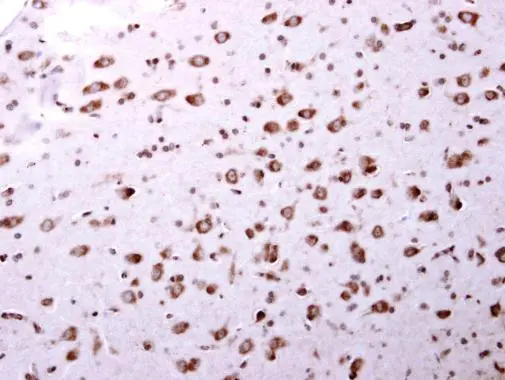
ARA9 antibody detects ARA9 protein at cytosol on mouse middle brain by immunohistochemical analysis. Sample: Paraffin-embedded mouse middle brain. ARA9 antibody (GTX113891) dilution: 1:500.
Antigen Retrieval: Trilogy? (EDTA based, pH 8.0) buffer, 15min
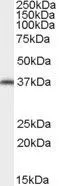
 A: Molt-4 (GTX27912) 10% SDS PAGE GTX113891 diluted at 1:1000")
![ARA9 antibody detects AIP protein at cytoplasm by confocal immunofluorescent analysis. Sample: HeLa cells were fixed in 4% paraformaldehyde at RT for 15 min. Green: AIP protein stained by ARA9 antibody (GTX113891) diluted at 1:500. Blue: Hoechst 33343 staining. [Images captured by Olympus FV10i Confocal Laser Scanning Microscope]](https://www.genetex.com/upload/website/prouct_img/normal/GTX113891/GTX113891_40150_IFA_w_23060501_503.webp "ARA9 antibody detects AIP protein at cytoplasm by confocal immunofluorescent analysis. Sample: HeLa cells were fixed in 4% paraformaldehyde at RT for 15 min. Green: AIP protein stained by ARA9 antibody (GTX113891) diluted at 1:500. Blue: Hoechst 33343 staining. [Images captured by Olympus FV10i Confocal Laser Scanning Microscope]")
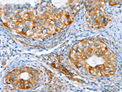
ARA9 antibody detects ARA9 protein at cytosol on mouse middle brain by immunohistochemical analysis. Sample: Paraffin-embedded mouse middle brain. ARA9 antibody (GTX113891) dilution: 1:500.
Antigen Retrieval: Trilogy? (EDTA based, pH 8.0) buffer, 15min
ARA9 antibody
GTX113891
ApplicationsImmunoFluorescence, Western Blot, ImmunoCytoChemistry, ImmunoHistoChemistry, ImmunoHistoChemistry Paraffin
Product group Antibodies
ReactivityHuman, Mouse
TargetAIP
Overview
- SupplierGeneTex
- Product NameARA9 antibody
- Delivery Days Customer9
- Application Supplier NoteWB: 1:500-1:3000. ICC/IF: 1:100-1:1000. IHC-P: 1:100-1:1000. *Optimal dilutions/concentrations should be determined by the researcher.Not tested in other applications.
- ApplicationsImmunoFluorescence, Western Blot, ImmunoCytoChemistry, ImmunoHistoChemistry, ImmunoHistoChemistry Paraffin
- CertificationResearch Use Only
- ClonalityPolyclonal
- Concentration1.01 mg/ml
- ConjugateUnconjugated
- Gene ID9049
- Target nameAIP
- Target descriptionAHR interacting HSP90 co-chaperone
- Target synonymsARA9, FKBP16, FKBP37, PITA1, SMTPHN, XAP-2, XAP2, AH receptor-interacting protein, Ah receptor activated 9, FK506-binding protein 37, FKBP prolyl isomerase 16, HBV X-associated protein 2, X-associated protein-2, aryl hydrocarbon receptor interacting protein, aryl hydrocarbon receptor-associated protein 9, hepatitis B virus X-associated cellular protein 2, immunophilin homolog ARA9
- HostRabbit
- IsotypeIgG
- Protein IDO00170
- Protein NameAH receptor-interacting protein
- Scientific DescriptionThe protein encoded by this gene is a receptor for aryl hydrocarbons and a ligand-activated transcription factor. The encoded protein is found in the cytoplasm as part of a multiprotein complex, but upon binding of ligand is transported to the nucleus. This protein can regulate the expression of many xenobiotic metabolizing enzymes. Also, the encoded protein can bind specifically to and inhibit the activity of hepatitis B virus. [provided by RefSeq]
- ReactivityHuman, Mouse
- Storage Instruction-20°C or -80°C,2°C to 8°C
- UNSPSC41116161
Datasheet
Related products
Product group Antibodies
Anti-AIP Antibody144-12546
ApplicationsWestern Blot
ReactivityHuman, Mouse, Rat
TargetAIP
- SizePrice
Product group Antibodies

ARA9 AntibodyABX430004
ApplicationsWestern Blot, ELISA, ImmunoHistoChemistry
- SizePrice
Product group Antibodies

Goat anti-AIP / ARA9EB07776
ApplicationsWestern Blot, ELISA, ImmunoHistoChemistry
ReactivityCanine, Human, Mouse, Rat
TargetAIP
- SizePrice
Product group Antibodies
AIP AntibodyCSB-PA049334
ApplicationsELISA, ImmunoHistoChemistry
ReactivityHuman, Mouse, Rat
TargetAIP
- SizePrice

Product group Antibodies
Anti-AIP AntibodyHPA050217
ApplicationsImmunoCytoChemistry
ReactivityHuman
TargetAIP
- SizePrice
Product group Antibodies

ARA9 / AIP AntibodyLS-C406135
ApplicationsELISA, ImmunoHistoChemistry
ReactivityHuman, Mouse, Rat
TargetAIP
- SizePrice
Product group Antibodies
ApplicationsImmunoPrecipitation, Western Blot, ImmunoCytoChemistry, ImmunoHistoChemistry
ReactivityMouse, Rat
TargetAIP
- SizePrice

Product group Antibodies
Anti-ARA9/AIP Antibody Picoband(r)PB9042-CARRIER-FREE
ApplicationsFlow Cytometry, ImmunoFluorescence, Western Blot, ImmunoCytoChemistry
ReactivityHuman
TargetAIP
- SizePrice
![ARA9 antibody [N1C3] detects ARA9 protein by Western blot analysis. A. 30 μg HeLa whole cell lysate/extract B. 30 μg Molt-4 whole cell lysate/extract C. 30 μg Raji whole cell lysate/extract 10 % SDS-PAGE ARA9 antibody [N1C3] (GTX110665) dilution: 1:5000](https://www.genetex.com/upload/website/prouct_img/normal/GTX110665/GTX110665_40044_WB_w_23060500_791.webp)
Product group Antibodies
ARA9 antibody [N1C3]GTX110665
ApplicationsImmunoFluorescence, Western Blot, ImmunoCytoChemistry, ImmunoHistoChemistry, ImmunoHistoChemistry Paraffin
ReactivityHuman, Mouse, Rat
TargetAIP
- SizePrice
Product group Antibodies
ARA9 antibody, C-termGTX89023
ApplicationsWestern Blot, ImmunoHistoChemistry, ImmunoHistoChemistry Paraffin
ReactivityHuman, Mouse
TargetAIP
- SizePrice