

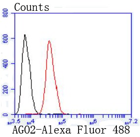
Flow cytometric analysis of Jurkat cells with Argonaute 2 (7C3) Monoclonal Antibody (bsm-52820R) at 1:50 dilution (red) compared with an unlabeled control (cells without incubation with primary antibody; black)
 Monoclonal Antibody (bsm-52820R) at 1:1000 overnight at 4°C followed by a conjugated secondary antibody for 60 minutes at 37°C.")
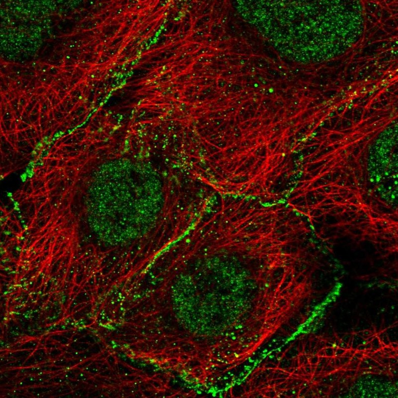
 staining with Argonaute 2 (7C3) Monoclonal Antibody (bsm-52820R) at 1:100 in AGS cells (red). The nuclear counterstain is DAPI (blue). Cells were fixed in paraformaldehyde, permeabilized with 0.25% Triton X100/PBS.")
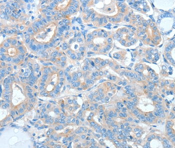
 Monoclonal Antibody (bsm-52820R) at 1:100, overnight at 4°C, followed by a conjugated secondary antibody and DAB staining. Counterstained with hematoxylin.")
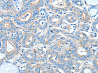
 Monoclonal Antibody (bsm-52820R) at 1:100, overnight at 4°C, followed by a conjugated secondary antibody and DAB staining. Counterstained with hematoxylin.")
Flow cytometric analysis of Jurkat cells with Argonaute 2 (7C3) Monoclonal Antibody (bsm-52820R) at 1:50 dilution (red) compared with an unlabeled control (cells without incubation with primary antibody; black)
AGO2 Recombinant Antibody
BSM-52820R
ApplicationsFlow Cytometry
Product group Antibodies
ReactivityHuman, Mouse, Rat
TargetAGO2
Overview
- SupplierBioss
- Product NameAGO2 Recombinant Antibody
- Delivery Days Customer16
- ApplicationsFlow Cytometry
- Applications SupplierFCM(1:20-100)
- CertificationResearch Use Only
- ClonalityMonoclonal
- ConjugateUnconjugated
- Gene ID27161
- Target nameAGO2
- Target descriptionargonaute RISC catalytic component 2
- Target synonymsCASC7, EIF2C2, LESKRES, LINC00980, PPD, Q10, protein argonaute-2, PAZ Piwi domain protein, argonaute 2, RISC catalytic component, cancer susceptibility candidate 7 (non-protein coding), eukaryotic translation initiation factor 2C, 2, long intergenic non-protein coding RNA 980, protein slicer
- HostRabbit
- IsotypeIgG
- Protein IDQ9UKV8
- Protein NameProtein argonaute-2
- ReactivityHuman, Mouse, Rat
- Storage Instruction-20°C
- UNSPSC41116161
Datasheet
Related products
Product group Antibodies
Anti-AGO2 AntibodyA46743
ApplicationsImmunoHistoChemistry
ReactivityHuman, Mouse
- SizePrice
Product group Antibodies
Anti-AGO2 Antibody144-06023
ApplicationsWestern Blot, ImmunoHistoChemistry
ReactivityHuman, Mouse, Rat
TargetAGO2
- SizePrice
Product group Antibodies
AGO2 AntibodyCSB-PA290707
ApplicationsELISA, ImmunoHistoChemistry
ReactivityHuman, Mouse, Rat
TargetAGO2
- SizePrice
Product group Antibodies
Ago2 Polyclonal AntibodyCAC07145
ApplicationsWestern Blot, ChIP Chromatin ImmunoPrecipitation, ELISA, ImmunoHistoChemistry
ReactivityMouse
TargetAGO2
- SizePrice
Product group Antibodies

AGO2 / EIF2C2 AntibodyLS-C483260
ApplicationsImmunoPrecipitation, ImmunoHistoChemistry, ImmunoHistoChemistry Paraffin
ReactivityMouse
TargetAGO2
- SizePrice
Product group Antibodies
Anti-AGO2 AntibodyHPA058075
ApplicationsImmunoCytoChemistry
ReactivityHuman
TargetAGO2
- SizePrice
Product group Antibodies
EIF2C2 antibody [C1C3]GTX117699
ApplicationsWestern Blot
ReactivityHuman
TargetAGO2
- SizePrice
Product group Antibodies
Anti-Argonaute-2 AntibodyCAB6023
ApplicationsWestern Blot, ELISA, ImmunoHistoChemistry, ImmunoHistoChemistry Paraffin
ReactivityHuman
TargetAGO2
- SizePrice

Product group Antibodies
Anti-Ago2/eIF2C2 Antibody Picoband(r)PB9978-CARRIER-FREE
ApplicationsWestern Blot
ReactivityBovine, Canine, Equine, Hamster, Human, Monkey, Mouse, Rabbit, Rat
TargetAGO2
- SizePrice