

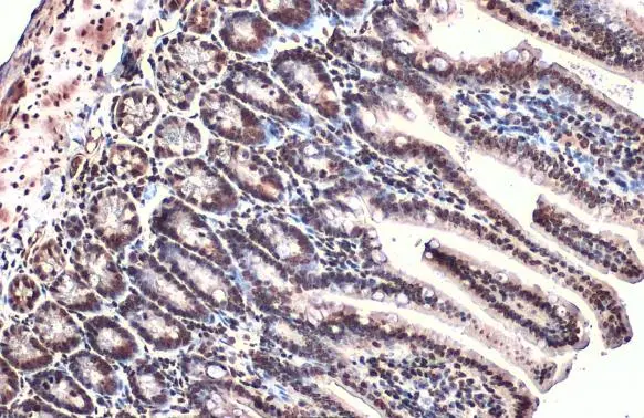
ARID5B antibody detects ARID5B protein at nucleus by immunohistochemical analysis. Sample: Paraffin-embedded mouse intestine. ARID5B stained by ARID5B antibody (GTX131249) diluted at 1:500. Antigen Retrieval: Citrate buffer, pH 6.0, 15 min
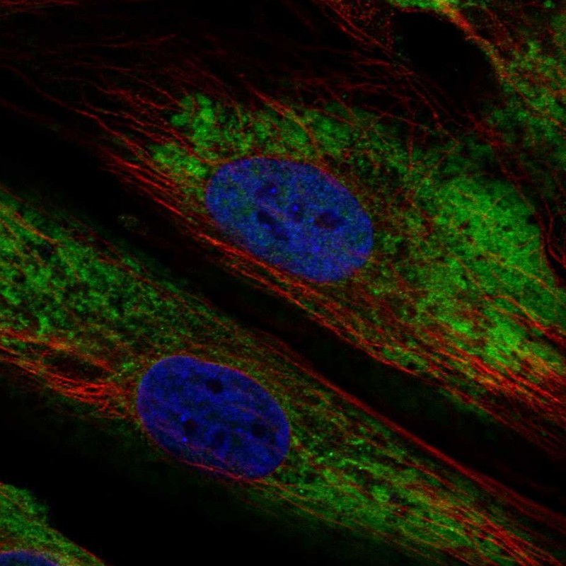
![ARID5B antibody detects ARID5B protein at nucleus by immunofluorescent analysis. Sample: HeLa cells were fixed in 4% paraformaldehyde at RT for 15 min. Green: ARID5B stained by ARID5B antibody (GTX131249) diluted at 1:500. Red: alpha Tubulin, a cytoskeleton marker, stained by alpha Tubulin antibody [GT114] (GTX628802) diluted at 1:1000. Blue: Fluoroshield with DAPI (GTX30920).](https://www.genetex.com/upload/website/prouct_img/normal/GTX131249/GTX131249_44160_20211126_ICC_IF_23041023_480.webp "ARID5B antibody detects ARID5B protein at nucleus by immunofluorescent analysis. Sample: HeLa cells were fixed in 4% paraformaldehyde at RT for 15 min. Green: ARID5B stained by ARID5B antibody (GTX131249) diluted at 1:500. Red: alpha Tubulin, a cytoskeleton marker, stained by alpha Tubulin antibody [GT114] (GTX628802) diluted at 1:1000. Blue: Fluoroshield with DAPI (GTX30920).")
 diluted at 1:500. Antigen Retrieval: Citrate buffer, pH 6.0, 15 min")
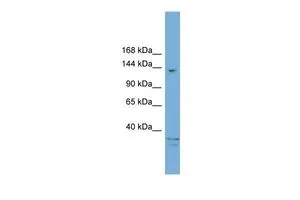
 were separated by 5% SDS-PAGE, and the membrane was blotted with ARID5B antibody (GTX131249) diluted at 1:1000. The HRP-conjugated anti-rabbit IgG antibody (GTX213110-01) was used to detect the primary antibody.")
ARID5B antibody detects ARID5B protein at nucleus by immunohistochemical analysis. Sample: Paraffin-embedded mouse intestine. ARID5B stained by ARID5B antibody (GTX131249) diluted at 1:500. Antigen Retrieval: Citrate buffer, pH 6.0, 15 min
ARID5B antibody
GTX131249
ApplicationsImmunoFluorescence, Western Blot, ImmunoCytoChemistry, ImmunoHistoChemistry, ImmunoHistoChemistry Paraffin
Product group Antibodies
ReactivityHuman, Mouse
TargetARID5B
Overview
- SupplierGeneTex
- Product NameARID5B antibody
- Delivery Days Customer9
- Application Supplier NoteWB: 1:500-1:3000. *Optimal dilutions/concentrations should be determined by the researcher.Not tested in other applications.
- ApplicationsImmunoFluorescence, Western Blot, ImmunoCytoChemistry, ImmunoHistoChemistry, ImmunoHistoChemistry Paraffin
- CertificationResearch Use Only
- ClonalityPolyclonal
- Concentration0.89 mg/ml
- ConjugateUnconjugated
- Gene ID84159
- Target nameARID5B
- Target descriptionAT-rich interaction domain 5B
- Target synonymsDESRT, MRF-2, MRF2, AT-rich interactive domain-containing protein 5B, ARID domain-containing protein 5B, AT-rich interactive domain 5B (MRF1-like), MRF1-like protein, modulator recognition factor 2 (MRF2)
- HostRabbit
- IsotypeIgG
- Protein IDQ14865
- Protein NameAT-rich interactive domain-containing protein 5B
- Scientific DescriptionThis gene encodes a member of the AT-rich interaction domain (ARID) family of DNA binding proteins. The encoded protein forms a histone H3K9Me2 demethylase complex with PHD finger protein 2 and regulates the transcription of target genes involved in adipogenesis and liver development. This gene also plays a role in cell growth and differentiation of B-lymphocyte progenitors, and single nucleotide polymorphisms in this gene are associated with acute lymphoblastic leukemia. Alternatively spliced transcript variants encoding multiple isoforms have been observed for this gene. [provided by RefSeq, Sep 2011]
- ReactivityHuman, Mouse
- Storage Instruction-20°C or -80°C,2°C to 8°C
- UNSPSC41116161
Datasheet
Related products

Product group Antibodies
ApplicationsWestern Blot
ReactivityHuman
TargetARID5B
- SizePrice
Product group Antibodies

ARID5B AntibodyLS-C783459
ApplicationsWestern Blot
ReactivityHuman
TargetARID5B
- SizePrice
Product group Antibodies
Anti-ARID5B AntibodyHPA015037
ApplicationsImmunoCytoChemistry
ReactivityHuman
TargetARID5B
- SizePrice
Product group Antibodies
ARID5B antibody, C-termGTX48904
ApplicationsWestern Blot
ReactivityHuman
TargetARID5B
- SizePrice