

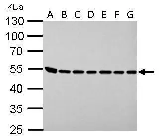
ASL antibody detects ASL protein by western blot analysis. A. 30 μg Neuro2A whole lysate/extract B. 30 μg GL261 whole cell lysate/extract C. 30 μg C8D30 whole cell lysate/extract D. 30 μg NIH-3T3 whole cell lysate/extract E. 30 μg BCL-1 whole cell lysate/extract F. 30 μg Raw264.7 whole cell lysate/extract G. 30 μg C2C12 whole cell lysate/extract 10% SDS-PAGE ASL antibody (GTX113629) dilution: 1:1000 The HRP-conjugated anti-rabbit IgG antibody (GTX213110-01) was used to detect the primary antibody.
 antibody at 1:500 dilution.
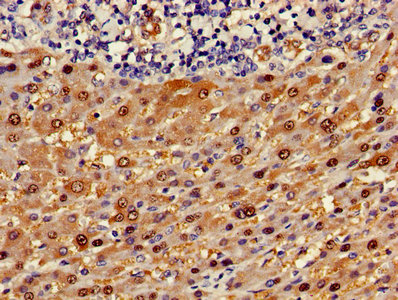
Antigen Retrieval: Trilogy? (EDTA based, pH 8.0) buffer, 15min")
 antibody at 1:200 dilution.")
 antibody at 1:500 dilution.
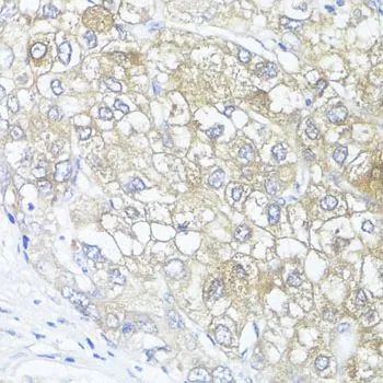
Antigen Retrieval: Trilogy? (EDTA based, pH 8.0) buffer, 15min")
 diluted at 1:500. Antigen Retrieval: Citrate buffer, pH 6.0, 15 min")
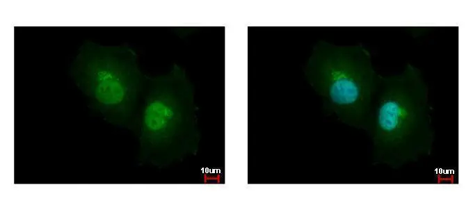
![ASL antibody detects ASL protein at cytoplasm by immunofluorescent analysis. Sample: HeLa cells were fixed in 4% paraformaldehyde at RT for 15 min. Green: ASL protein stained by ASL antibody (GTX113629) diluted at 1:500. Red: alpha Tubulin, a cytoskeleton marker, stained by alpha Tubulin antibody [GT114] (GTX628802) diluted at 1:1000. Blue: Hoechst 33342 staining.](https://www.genetex.com/upload/website/prouct_img/normal/GTX113629/GTX113629_40142_20150410_IFA_w_23060501_366.webp "ASL antibody detects ASL protein at cytoplasm by immunofluorescent analysis. Sample: HeLa cells were fixed in 4% paraformaldehyde at RT for 15 min. Green: ASL protein stained by ASL antibody (GTX113629) diluted at 1:500. Red: alpha Tubulin, a cytoskeleton marker, stained by alpha Tubulin antibody [GT114] (GTX628802) diluted at 1:1000. Blue: Hoechst 33342 staining.")
 were separated by 10% SDS-PAGE, and the membrane was blotted with ASL antibody (GTX113629) diluted at 1:1000. The HRP-conjugated anti-rabbit IgG antibody (GTX213110-01) was used to detect the primary antibody. Corresponding RNA expression data for the same cell lines are based on Human Protein Atlas program.")
ASL antibody detects ASL protein by western blot analysis. A. 30 μg Neuro2A whole lysate/extract B. 30 μg GL261 whole cell lysate/extract C. 30 μg C8D30 whole cell lysate/extract D. 30 μg NIH-3T3 whole cell lysate/extract E. 30 μg BCL-1 whole cell lysate/extract F. 30 μg Raw264.7 whole cell lysate/extract G. 30 μg C2C12 whole cell lysate/extract 10% SDS-PAGE ASL antibody (GTX113629) dilution: 1:1000 The HRP-conjugated anti-rabbit IgG antibody (GTX213110-01) was used to detect the primary antibody.
ASL antibody
GTX113629
ApplicationsImmunoFluorescence, Western Blot, ImmunoCytoChemistry, ImmunoHistoChemistry, ImmunoHistoChemistry Paraffin
Product group Antibodies
ReactivityHuman, Mouse
TargetASL
Overview
- SupplierGeneTex
- Product NameASL antibody
- Delivery Days Customer9
- Application Supplier NoteWB: 1:500-1:3000. ICC/IF: 1:100-1:1000. IHC-P: 1:100-1:1000. *Optimal dilutions/concentrations should be determined by the researcher.Not tested in other applications.
- ApplicationsImmunoFluorescence, Western Blot, ImmunoCytoChemistry, ImmunoHistoChemistry, ImmunoHistoChemistry Paraffin
- CertificationResearch Use Only
- ClonalityPolyclonal
- Concentration0.78 mg/ml
- ConjugateUnconjugated
- Gene ID435
- Target nameASL
- Target descriptionargininosuccinate lyase
- Target synonymsASAL, ASLD, argininosuccinate lyase, argininosuccinase, arginosuccinase
- HostRabbit
- IsotypeIgG
- Protein IDP04424
- Protein NameArgininosuccinate lyase
- Scientific DescriptionThis gene encodes a member of the lyase 1 family. The encoded protein forms a cytosolic homotetramer and primarily catalyzes the reversible hydrolytic cleavage of argininosuccinate into arginine and fumarate, an essential step in the liver in detoxifying ammonia via the urea cycle. Mutations in this gene result in the autosomal recessive disorder argininosuccinic aciduria, or argininosuccinic acid lyase deficiency. A nontranscribed pseudogene is also located on the long arm of chromosome 22. Alternatively spliced transcript variants encoding different isoforms have been described. [provided by RefSeq]
- ReactivityHuman, Mouse
- Storage Instruction-20°C or -80°C,2°C to 8°C
- UNSPSC41116161
Datasheet
Related products

Product group Antibodies
Anti-ASL AntibodyA31320
ApplicationsWestern Blot, ImmunoHistoChemistry
ReactivityHuman, Mouse
- SizePrice

Product group Antibodies
Anti-Argininosuccinate Lyase/ASL Antibody Picoband(r)A00742-1-CARRIER-FREE
ApplicationsWestern Blot
ReactivityMouse, Rat
TargetASL
- SizePrice
Product group Antibodies
Anti-ASL Antibody107-10865
ApplicationsImmunoFluorescence, Western Blot, ImmunoCytoChemistry, ImmunoHistoChemistry, ImmunoHistoChemistry Paraffin
ReactivityHuman
TargetASL
- SizePrice
Product group Antibodies
ApplicationsImmunoFluorescence, Western Blot, ELISA, ImmunoCytoChemistry, ImmunoHistoChemistry, ImmunoHistoChemistry Frozen, ImmunoHistoChemistry Paraffin
ReactivityCanine, Equine, Human, Mouse, Porcine, Rat
TargetASL
- SizePrice
Product group Antibodies
ASL AntibodyCSB-PA002213LA01HU
ApplicationsImmunoFluorescence, ELISA, ImmunoHistoChemistry
ReactivityHuman
TargetASL
- SizePrice
Product group Antibodies
Asl Polyclonal AntibodyCAC11756
ApplicationsImmunoFluorescence, ELISA, ImmunoHistoChemistry
TargetASL
- SizePrice
Product group Antibodies

ASL / Argininosuccinate Lyase AntibodyLS-C404547
ApplicationsELISA, ImmunoHistoChemistry
ReactivityHuman, Mouse, Rat
TargetASL
- SizePrice
Product group Antibodies
ASL antibodyGTX109750
ApplicationsImmunoFluorescence, Western Blot, ImmunoCytoChemistry, ImmunoHistoChemistry, ImmunoHistoChemistry Paraffin
ReactivityHuman
TargetASL
- SizePrice
Product group Antibodies
ASL antibodyGTX55520
ApplicationsImmunoFluorescence, Western Blot, ImmunoCytoChemistry, ImmunoHistoChemistry, ImmunoHistoChemistry Paraffin
ReactivityHuman, Mouse, Rat
TargetASL
- SizePrice

Product group Antibodies
Anti-ASL AntibodyHPA016646
ApplicationsImmunoHistoChemistry
ReactivityHuman
TargetASL
- SizePrice