

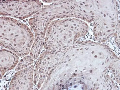
Immunohistochemical analysis of paraffin-embedded Cal27 xenograft, using Aspartoacylase(GTX113389) antibody at 1:100 dilution.
Antigen Retrieval: Trilogy? (EDTA based, pH 8.0) buffer, 15min
![Non-transfected (–) and transfected (+) 293T whole cell extracts (30 μg) were separated by 10% SDS-PAGE, and the membrane was blotted with Aspartoacylase antibody [N1C3-2] (GTX113389) diluted at 1:10000. The HRP-conjugated anti-rabbit IgG antibody (GTX213110-01) was used to detect the primary antibody.](https://www.genetex.com/upload/website/prouct_img/normal/GTX113389/GTX113389_40128_20180914_WB_B_w_23060501_848.webp "Non-transfected (–) and transfected (+) 293T whole cell extracts (30 μg) were separated by 10% SDS-PAGE, and the membrane was blotted with Aspartoacylase antibody [N1C3-2] (GTX113389) diluted at 1:10000. The HRP-conjugated anti-rabbit IgG antibody (GTX213110-01) was used to detect the primary antibody.")
![Various tissue extracts (50 μg) were separated by 10% SDS-PAGE, and the membrane was blotted with Aspartoacylase antibody [N1C3-2] (GTX113389) diluted at 1:1000. The HRP-conjugated anti-rabbit IgG antibody (GTX213110-01) was used to detect the primary antibody.](https://www.genetex.com/upload/website/prouct_img/normal/GTX113389/GTX113389_43824_20200117_WB_M_tissue_w_23060501_493.webp "Various tissue extracts (50 μg) were separated by 10% SDS-PAGE, and the membrane was blotted with Aspartoacylase antibody [N1C3-2] (GTX113389) diluted at 1:1000. The HRP-conjugated anti-rabbit IgG antibody (GTX213110-01) was used to detect the primary antibody.")
![Various whole cell extracts (30 μg) were separated by 12% SDS-PAGE, and the membrane was blotted with Aspartoacylase antibody [N1C3-2] (GTX113389) diluted at 1:1000. The HRP-conjugated anti-rabbit IgG antibody (GTX213110-01) was used to detect the primary antibody.](https://www.genetex.com/upload/website/prouct_img/normal/GTX113389/GTX113389_43824_20200131_WB_w_23060501_892.webp "Various whole cell extracts (30 μg) were separated by 12% SDS-PAGE, and the membrane was blotted with Aspartoacylase antibody [N1C3-2] (GTX113389) diluted at 1:1000. The HRP-conjugated anti-rabbit IgG antibody (GTX213110-01) was used to detect the primary antibody.")
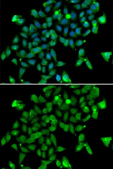
![Aspartoacylase antibody [N1C3-2] detects Aspartoacylase protein at cytoplasm by immunofluorescent analysis. Sample: HeLa cells were fixed in 4% paraformaldehyde at RT for 15 min. Green: Aspartoacylase protein stained by Aspartoacylase antibody [N1C3-2] (GTX113389) diluted at 1:500. Red: alpha Tubulin, a cytoskeleton marker, stained by alpha Tubulin antibody [GT114] (GTX628802) diluted at 1:1000. Blue: Hoechst 33342 staining.](https://www.genetex.com/upload/website/prouct_img/normal/GTX113389/GTX113389_40128_20150410_IFA_w_23060501_372.webp "Aspartoacylase antibody [N1C3-2] detects Aspartoacylase protein at cytoplasm by immunofluorescent analysis. Sample: HeLa cells were fixed in 4% paraformaldehyde at RT for 15 min. Green: Aspartoacylase protein stained by Aspartoacylase antibody [N1C3-2] (GTX113389) diluted at 1:500. Red: alpha Tubulin, a cytoskeleton marker, stained by alpha Tubulin antibody [GT114] (GTX628802) diluted at 1:1000. Blue: Hoechst 33342 staining.")
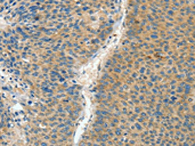
Immunohistochemical analysis of paraffin-embedded Cal27 xenograft, using Aspartoacylase(GTX113389) antibody at 1:100 dilution.
Antigen Retrieval: Trilogy? (EDTA based, pH 8.0) buffer, 15min
Aspartoacylase antibody [N1C3-2]
GTX113389
ApplicationsImmunoFluorescence, Western Blot, ImmunoCytoChemistry, ImmunoHistoChemistry, ImmunoHistoChemistry Paraffin
Product group Antibodies
ReactivityHuman, Monkey, Mouse
TargetASPA
Overview
- SupplierGeneTex
- Product NameAspartoacylase antibody [N1C3-2]
- Delivery Days Customer9
- Application Supplier NoteWB: 1:5000-1:20000. ICC/IF: 1:100-1:1000. IHC-P: 1:100-1:1000. *Optimal dilutions/concentrations should be determined by the researcher.Not tested in other applications.
- ApplicationsImmunoFluorescence, Western Blot, ImmunoCytoChemistry, ImmunoHistoChemistry, ImmunoHistoChemistry Paraffin
- CertificationResearch Use Only
- ClonalityPolyclonal
- Concentration1.33 mg/ml
- ConjugateUnconjugated
- Gene ID443
- Target nameASPA
- Target descriptionaspartoacylase
- Target synonymsACY2, ASP, aspartoacylase, ACY-2, aminoacylase 2, cytosolic aspartoacylase
- HostRabbit
- IsotypeIgG
- Protein IDP45381
- Protein NameAspartoacylase
- Scientific DescriptionThis gene encodes an enzyme that catalyzes the conversion of N-acetyl_L-aspartic acid (NAA) to aspartate and acetate. NAA is abundant in the brain where hydrolysis by aspartoacylase is thought to help maintain white matter. This protein is an NAA scavenger in other tissues. Mutations in this gene cause Canavan disease. Alternatively spliced transcript variants have been found for this gene. [provided by RefSeq]
- ReactivityHuman, Monkey, Mouse
- Storage Instruction-20°C or -80°C,2°C to 8°C
- UNSPSC41116161
Datasheet
Related products
Product group Antibodies
ApplicationsImmunoFluorescence, Western Blot, ImmunoCytoChemistry
ReactivityHuman, Mouse, Rat
TargetASPA
- SizePrice
Product group Antibodies
Anti-ASPA AntibodyA31964
ApplicationsImmunoFluorescence, Western Blot, ImmunoHistoChemistry
ReactivityHuman
- SizePrice
Product group Antibodies
Anti-ASPA Antibody144-07271
ApplicationsImmunoFluorescence, Western Blot
ReactivityHuman, Mouse, Rat
TargetASPA
- SizePrice
Product group Antibodies
ASPA AntibodyCSB-PA075666
ApplicationsWestern Blot, ELISA, ImmunoHistoChemistry
ReactivityHuman, Mouse, Rat
TargetASPA
- SizePrice
Product group Antibodies

ASPA AntibodyLS-C401413
ApplicationsWestern Blot, ELISA, ImmunoHistoChemistry
ReactivityHuman, Mouse, Rat
TargetASPA
- SizePrice

Product group Antibodies
Anti-ASPA-25ulHPA022142
ApplicationsWestern Blot, ImmunoHistoChemistry
ReactivityHuman
- SizePrice
Product group Antibodies
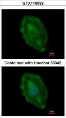
Aspartoacylase antibodyGTX110699
ApplicationsImmunoFluorescence, Western Blot, ImmunoCytoChemistry
ReactivityHuman, Mouse
TargetASPA
- SizePrice
Product group Antibodies
Aspartoacylase antibodyGTX55521
ApplicationsImmunoFluorescence, Western Blot, ImmunoCytoChemistry
ReactivityHuman, Mouse, Rat
TargetASPA
- SizePrice
Product group Antibodies
Anti-ASPA AntibodyCAB7271
ApplicationsImmunoFluorescence, Western Blot, ELISA, ImmunoCytoChemistry
ReactivityHuman
TargetASPA
- SizePrice