

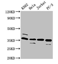
Western Blot Positive WB detected in: K562 whole cell lysate, Hela whole cell lysate, Jurkat whole cell lysate, PC-3 whole cell lysate All lanes: ATG3 antibody at 4microg/ml Secondary Goat polyclonal to rabbit IgG at 1/50000 dilution Predicted band size: 36 kDa Observed band size: 36 kDa
. Section was blocked with 10% normal goat serum 30min at RT. Then primary antibody (1% BSA) was incubated at 4°C overnight. The primary is detected by a biotinylated secondary antibody and visualized using an HRP conjugated SP system.")
. Section was blocked with 10% normal goat serum 30min at RT. Then primary antibody (1% BSA) was incubated at 4°C overnight. The primary is detected by a biotinylated secondary antibody and visualized using an HRP conjugated SP system.")
.")
Western Blot Positive WB detected in: K562 whole cell lysate, Hela whole cell lysate, Jurkat whole cell lysate, PC-3 whole cell lysate All lanes: ATG3 antibody at 4microg/ml Secondary Goat polyclonal to rabbit IgG at 1/50000 dilution Predicted band size: 36 kDa Observed band size: 36 kDa
ATG3 Antibody
CSB-PA002288HA01HU
ApplicationsImmunoFluorescence, Western Blot, ELISA, ImmunoHistoChemistry
Product group Antibodies
ReactivityHuman
TargetATG3
Overview
- SupplierCusabio
- Product NameATG3 Antibody
- Delivery Days Customer20
- ApplicationsImmunoFluorescence, Western Blot, ELISA, ImmunoHistoChemistry
- CertificationResearch Use Only
- ClonalityPolyclonal
- ConjugateUnconjugated
- Gene ID64422
- Target nameATG3
- Target descriptionautophagy related 3
- Target synonymsAPG3, APG3-LIKE, APG3L, PC3-96, hApg3, ubiquitin-like-conjugating enzyme ATG3, APG3 autophagy 3-like, ATG3 autophagy related 3 homolog, autophagy-related protein 3
- HostRabbit
- IsotypeIgG
- Protein IDQ9NT62
- Protein NameUbiquitin-like-conjugating enzyme ATG3
- Scientific DescriptionE2 conjugating enzyme required for the cytoplasm to vacuole transport (Cvt), autophagy, and mitochondrial homeostasis. Responsible for the E2-like covalent binding of phosphatidylethanolamine to the C-terminal Gly of ATG8-like proteins (GABARAP, GABARAPL1, GABARAPL2 or MAP1LC3A). The ATG12-ATG5 conjugate plays a role of an E3 and promotes the transfer of ATG8-like proteins from ATG3 to phosphatidylethanolamine (PE). This step is required for the membrane association of ATG8-like proteins. The formation of the ATG8-phosphatidylethanolamine conjugates is essential for autophagy and for the cytoplasm to vacuole transport (Cvt). Preferred substrate is MAP1LC3A. Also acts as an autocatalytic E2-like enzyme, catalyzing the conjugation of ATG12 to itself, ATG12 conjugation to ATG3 playing a role in mitochondrial homeostasis but not in autophagy. ATG7 (E1-like enzyme) facilitates this reaction by forming an E1-E2 complex with ATG3. Promotes primary ciliogenesis by removing OFD1 from centriolar satellites via the autophagic pathway.
- ReactivityHuman
- Storage Instruction-20°C or -80°C
- UNSPSC41116161
Related products
Product group Antibodies
Anti-ATG3 AntibodyA31063
ApplicationsWestern Blot, ImmunoHistoChemistry
ReactivityHuman, Mouse, Rat
- SizePrice
Product group Antibodies
Anti-ATG3 Antibody144-05809
ApplicationsWestern Blot, ImmunoHistoChemistry
ReactivityHuman, Mouse
TargetATG3
- SizePrice

Product group Antibodies
Anti-Apg3/ATG3 Antibody Picoband(r)A01768-1-CARRIER-FREE
ApplicationsWestern Blot
ReactivityHuman, Monkey, Mouse
TargetATG3
- SizePrice
Product group Antibodies
ATG3 Recombinant Antibody, AbBy Fluor-488 ConjugatedBSM-61474R-BF488
ApplicationsWestern Blot
ReactivityHuman, Mouse, Rat
TargetATG3
- SizePrice
Product group Antibodies
Atg3 Polyclonal AntibodyCAC07035
ApplicationsImmunoFluorescence, Western Blot, ELISA, ImmunoHistoChemistry
TargetATG3
- SizePrice
Product group Antibodies

ATG3 AntibodyLS-C401261
ApplicationsELISA, ImmunoHistoChemistry
ReactivityHuman, Mouse, Rat
TargetATG3
- SizePrice

Product group Antibodies
Anti-ATG3 AntibodyHPA040471
ApplicationsImmunoHistoChemistry
ReactivityHuman
TargetATG3
- SizePrice
Product group Antibodies
ATG3 antibodyGTX128065
ApplicationsWestern Blot
ReactivityHuman, Mouse, Rat
TargetATG3
- SizePrice
Product group Antibodies
Anti-ATG3 AntibodyCAB5809
ApplicationsImmunoFluorescence, Western Blot, ELISA, ImmunoCytoChemistry
ReactivityHuman
TargetATG3
- SizePrice