

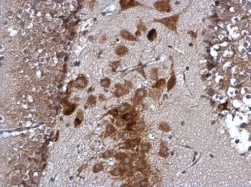
ATXN2 antibody detects ATXN2 protein at cytoplasm in mouse brain by immunohistochemical analysis. Sample: Paraffin-embedded mouse brain. ATXN2 antibody (GTX130329) diluted at 1:500.
Antigen Retrieval: Citrate buffer, pH 6.0, 15 min
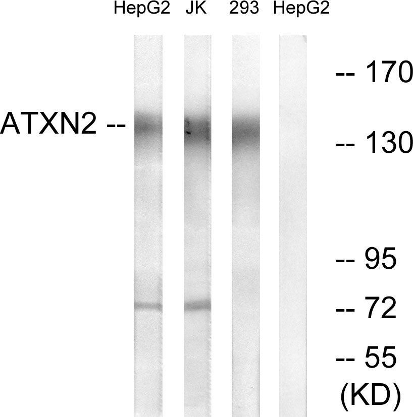
 and transfected (+) 293T whole cell extracts (30 μg) were separated by 5% SDS-PAGE, and the membrane was blotted with ATXN2 antibody (GTX130329) diluted at 1:3000. The HRP-conjugated anti-rabbit IgG antibody (GTX213110-01) was used to detect the primary antibody.")
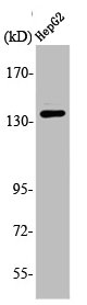
 were separated by 5% SDS-PAGE, and the membrane was blotted with ATXN2 antibody (GTX130329) diluted at 1:1000. The HRP-conjugated anti-rabbit IgG antibody (GTX213110-01) was used to detect the primary antibody.")
ATXN2 antibody detects ATXN2 protein at cytoplasm in mouse brain by immunohistochemical analysis. Sample: Paraffin-embedded mouse brain. ATXN2 antibody (GTX130329) diluted at 1:500.
Antigen Retrieval: Citrate buffer, pH 6.0, 15 min
ATXN2 antibody
GTX130329
ApplicationsImmunoPrecipitation, Western Blot, ImmunoHistoChemistry, ImmunoHistoChemistry Paraffin
Product group Antibodies
ReactivityHuman, Mouse
TargetATXN2
Overview
- SupplierGeneTex
- Product NameATXN2 antibody
- Delivery Days Customer9
- Application Supplier NoteWB: 1:500-1:3000. IHC-P: 1:100-1:1000. *Optimal dilutions/concentrations should be determined by the researcher.Not tested in other applications.
- ApplicationsImmunoPrecipitation, Western Blot, ImmunoHistoChemistry, ImmunoHistoChemistry Paraffin
- CertificationResearch Use Only
- ClonalityPolyclonal
- Concentration0.2 mg/ml
- ConjugateUnconjugated
- Gene ID6311
- Target nameATXN2
- Target descriptionataxin 2
- Target synonymsATX2, SCA2, TNRC13, ataxin-2, spinocerebellar ataxia type 2 protein, trinucleotide repeat-containing gene 13 protein
- HostRabbit
- IsotypeIgG
- Protein IDQ99700
- Protein NameAtaxin-2
- Scientific DescriptionThe autosomal dominant cerebellar ataxias (ADCA) are a heterogeneous group of neurodegenerative disorders characterized by progressive degeneration of the cerebellum, brain stem and spinal cord. Clinically, ADCA has been divided into three groups: ADCA types I-III. Defects in this gene are the cause of spinocerebellar ataxia type 2 (SCA2). SCA2 belongs to the autosomal dominant cerebellar ataxias type I (ADCA I) which are characterized by cerebellar ataxia in combination with additional clinical features like optic atrophy, ophthalmoplegia, bulbar and extrapyramidal signs, peripheral neuropathy and dementia. SCA2 is caused by expansion of a CAG repeat in the coding region of this gene. Longer expansions result in earlier onset of the disease. Alternatively spliced transcript variants encoding different isoforms have been identified but their full length sequence has not been determined. [provided by RefSeq]
- ReactivityHuman, Mouse
- Storage Instruction-20°C or -80°C,2°C to 8°C
- UNSPSC41116161
Datasheet
Related products
Product group Antibodies
ATXN2 AntibodyCSB-PA000944
ApplicationsWestern Blot, ELISA, ImmunoHistoChemistry
ReactivityHuman
TargetATXN2
- SizePrice

Product group Antibodies
Anti-SCA2/ATXN2 Antibody Picoband(r)A01915-CARRIER-FREE
ApplicationsWestern Blot, ELISA, ImmunoHistoChemistry
ReactivityHuman
TargetATXN2
- SizePrice
Product group Antibodies
Anti-ATXN2 AntibodyA101206
ApplicationsWestern Blot, ELISA
ReactivityHuman
- SizePrice
Product group Antibodies

Goat anti-ATXN2EB10217
ApplicationsFlow Cytometry, ImmunoFluorescence, ELISA
ReactivityBovine, Canine, Human, Mouse, Porcine
TargetATXN2
- SizePrice

Product group Antibodies
Anti-ATXN2 AntibodyHPA018295
ApplicationsImmunoCytoChemistry, ImmunoHistoChemistry
ReactivityHuman
TargetATXN2
- SizePrice
Product group Antibodies

ApplicationsELISA
ReactivityHuman
TargetATXN2
- SizePrice
Product group Antibodies
Atxn2 Polyclonal AntibodyCAC08114
ApplicationsImmunoFluorescence, Western Blot, ELISA
TargetATXN2
- SizePrice
![ATXN2 antibody [HL1902] detects ATXN2 protein at cytoplasm by immunofluorescent analysis. Sample: A431 cells were fixed in 4% paraformaldehyde at RT for 15 min. Green: ATXN2 stained by ATXN2 antibody [HL1902] (GTX637651) diluted at 1:500. Blue: Fluoroshield with DAPI (GTX30920).](https://www.genetex.com/upload/website/prouct_img/normal/GTX637651/GTX637651_T-44837_20230106_ICC_IF_23013122_562.webp)
Product group Antibodies
ATXN2 antibody [HL1902]GTX637651
ApplicationsImmunoFluorescence, Western Blot, ImmunoCytoChemistry, ImmunoHistoChemistry, ImmunoHistoChemistry Paraffin
ReactivityHuman, Rat
TargetATXN2
- SizePrice
![Whole cell extract (30 μg) was separated by 5% SDS-PAGE, and the membrane was blotted with ATXN2 antibody [HL1903] (GTX637652) diluted at 1:1000. The HRP-conjugated anti-rabbit IgG antibody (GTX213110-01) was used to detect the primary antibody.](https://www.genetex.com/upload/website/prouct_img/normal/GTX637652/GTX637652_T-44837_20221209_WB_22121123_552.webp)
Product group Antibodies
ATXN2 antibody [HL1903]GTX637652
ApplicationsWestern Blot
ReactivityHuman
TargetATXN2
- SizePrice
![Whole cell extract (30 μg) was separated by 5% SDS-PAGE, and the membrane was blotted with ATXN2 antibody [HL2885] (GTX640164) diluted at 1:1000. The HRP-conjugated anti-rabbit IgG antibody (GTX213110-01) was used to detect the primary antibody.](https://www.genetex.com/upload/website/prouct_img/normal/GTX640164/GTX640164_T-45369_20240503_WB_R_24050701_284.webp)
Product group Antibodies
ATXN2 antibody [HL2885]GTX640164
ApplicationsImmunoFluorescence, Western Blot, ImmunoCytoChemistry, ImmunoHistoChemistry, ImmunoHistoChemistry Paraffin
ReactivityHuman, Rat
TargetATXN2
- SizePrice