

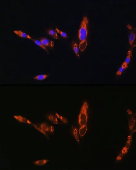
ICC/IF analysis of NIH/3T3 cells using GTX64617 AVPR1A antibody. Blue : DAPI Dilution : 1:100


ICC/IF analysis of NIH/3T3 cells using GTX64617 AVPR1A antibody. Blue : DAPI Dilution : 1:100
AVPR1A antibody
GTX64617
ApplicationsImmunoFluorescence, Western Blot, ImmunoCytoChemistry
Product group Antibodies
ReactivityHuman, Mouse, Rat
TargetAVPR1A
Overview
- SupplierGeneTex
- Product NameAVPR1A antibody
- Delivery Days Customer9
- Application Supplier NoteWB: 1:500 - 1:2000. ICC/IF: 1:50 - 1:200. *Optimal dilutions/concentrations should be determined by the researcher.Not tested in other applications.
- ApplicationsImmunoFluorescence, Western Blot, ImmunoCytoChemistry
- CertificationResearch Use Only
- ClonalityPolyclonal
- ConjugateUnconjugated
- Gene ID552
- Target nameAVPR1A
- Target descriptionarginine vasopressin receptor 1A
- Target synonymsAVPR V1a, AVPR1, V1aR, vasopressin V1a receptor, SCCL vasopressin subtype 1a receptor, V1-vascular vasopressin receptor AVPR1A, V1a vasopressin receptor, antidiuretic hormone receptor 1A, vascular/hepatic-type arginine vasopressin receptor
- HostRabbit
- IsotypeIgG
- Protein IDP37288
- Protein NameVasopressin V1a receptor
- Scientific DescriptionThe protein encoded by this gene acts as receptor for arginine vasopressin. This receptor belongs to the subfamily of G-protein coupled receptors which includes AVPR1B, V2R and OXT receptors. Its activity is mediated by G proteins which stimulate a phosphatidylinositol-calcium second messenger system. The receptor mediates cell contraction and proliferation, platelet aggregation, release of coagulation factor and glycogenolysis. [provided by RefSeq, Jul 2008]
- ReactivityHuman, Mouse, Rat
- Storage Instruction-20°C or -80°C,2°C to 8°C
- UNSPSC41116161
Datasheet
Related products
Product group Antibodies
AVPR1A AntibodyCSB-PA002468LA01HU
ApplicationsImmunoFluorescence, ELISA, ImmunoHistoChemistry
ReactivityHuman
TargetAVPR1A
- SizePrice
Product group Antibodies
AVPR1A Polyclonal AntibodyCAC12991
ApplicationsImmunoFluorescence, ELISA, ImmunoHistoChemistry
TargetAVPR1A
- SizePrice
Product group Antibodies
Anti-AVPR1A Antibody144-08400
ApplicationsWestern Blot
ReactivityHuman, Mouse, Rat
TargetAVPR1A
- SizePrice
Product group Antibodies
ApplicationsWestern Blot, ELISA
ReactivityHuman, Mouse, Rat
- SizePrice
Product group Antibodies
Anti-AVPR1A/V1aR Antibody Picoband(r)A03279-CARRIER-FREE
ApplicationsWestern Blot, ELISA
ReactivityHuman
TargetAVPR1A
- SizePrice
Product group Antibodies

Goat anti-AVPR1AEB07654
ApplicationsWestern Blot, ELISA
ReactivityBovine, Canine, Human, Mouse, Porcine, Rat
TargetAVPR1A
- SizePrice
Product group Antibodies

AVPR1A / V1a Receptor AntibodyLS-C409934
ApplicationsWestern Blot
ReactivityHuman, Mouse, Rat
TargetAVPR1A
- SizePrice
Product group Antibodies
References
AVPR1A antibody, InternalGTX89114
ApplicationsWestern Blot
ReactivityHuman, Mouse, Rodent, Rat
TargetAVPR1A
- SizePrice
Product group Antibodies
References
AVPR1A Polyclonal AntibodyBS-11598R
ApplicationsImmunoFluorescence, ImmunoPrecipitation, Western Blot, ELISA, ImmunoCytoChemistry, ImmunoHistoChemistry, ImmunoHistoChemistry Paraffin
ReactivityHuman, Mouse, Rabbit, Rat
TargetAVPR1A
- SizePrice