

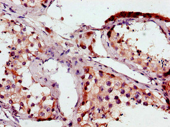
B-Raf antibody [N2C1], Internal detects B-Raf protein at cytosol on mouse testis by immunohistochemical analysis. Sample: Paraffin-embedded mouse testis. B-Raf antibody [N2C1], Internal (GTX100913) dilution: 1:500.
Antigen Retrieval: Trilogy? (EDTA based, pH 8.0) buffer, 15min
![B-Raf antibody [N2C1], Internal immunoprecipitates B-Raf protein in IP experiments.
IP Sample: HepG2 whole cell lysate/extract
A : 30 μg whole cell lysate/extract of B-Raf protein expressing HepG2 cells
B : Control with 3 μg of pre-immune rabbit IgG
C : Immunoprecipitation of B-Raf by 3 μg of B-Raf antibody [N2C1], Internal (GTX100913)
10% SDS-PAGE
The immunoprecipitated B-Raf protein was detected by B-Raf antibody [N2C1], Internal (GTX100913) diluted at 1:500.
EasyBlot anti-rabbit IgG (HRP) (GTX221666-01) was used as a secondary reagent.](https://www.genetex.com/upload/website/prouct_img/normal/GTX100913/GTX100913_39471_IP_w_23060100_499.webp "B-Raf antibody [N2C1], Internal immunoprecipitates B-Raf protein in IP experiments.
IP Sample: HepG2 whole cell lysate/extract
A : 30 μg whole cell lysate/extract of B-Raf protein expressing HepG2 cells
B : Control with 3 μg of pre-immune rabbit IgG
C : Immunoprecipitation of B-Raf by 3 μg of B-Raf antibody [N2C1], Internal (GTX100913)
10% SDS-PAGE
The immunoprecipitated B-Raf protein was detected by B-Raf antibody [N2C1], Internal (GTX100913) diluted at 1:500.
EasyBlot anti-rabbit IgG (HRP) (GTX221666-01) was used as a secondary reagent.")
![Various whole cell extracts (30 μg) were separated by 7.5% SDS-PAGE, and the membrane was blotted with B-Raf antibody [N2C1], Internal (GTX100913) diluted at 1:1000. The HRP-conjugated anti-rabbit IgG antibody (GTX213110-01) was used to detect the primary antibody.](https://www.genetex.com/upload/website/prouct_img/normal/GTX100913/GTX100913_43607_20190628_WB_w_23060100_350.webp "Various whole cell extracts (30 μg) were separated by 7.5% SDS-PAGE, and the membrane was blotted with B-Raf antibody [N2C1], Internal (GTX100913) diluted at 1:1000. The HRP-conjugated anti-rabbit IgG antibody (GTX213110-01) was used to detect the primary antibody.")
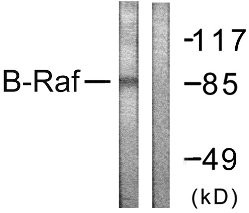
![B-Raf antibody [N2C1], Internal detects B-Raf protein by western blot analysis. Non-transfected (-) and B-Raf-transfected (+) 293T whole cell extracts (30 μg) were separated by 7.5% SDS-PAGE, and the membrane was blotted with B-Raf antibody [N2C1], Internal (GTX100913) diluted at 1:5000.](https://www.genetex.com/upload/website/prouct_img/normal/GTX100913/GTX100913_39471_20151029_WB_B_w_23060100_427.webp "B-Raf antibody [N2C1], Internal detects B-Raf protein by western blot analysis. Non-transfected (-) and B-Raf-transfected (+) 293T whole cell extracts (30 μg) were separated by 7.5% SDS-PAGE, and the membrane was blotted with B-Raf antibody [N2C1], Internal (GTX100913) diluted at 1:5000.")
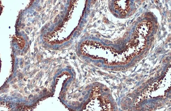
![B-Raf antibody [N2C1], Internal detects B-Raf protein at cytoplasm by immunohistochemical analysis. Sample: Paraffin-embedded human breast carcinoma. B-Raf stained by B-Raf antibody [N2C1], Internal (GTX100913) diluted at 1:500. Antigen Retrieval: Citrate buffer, pH 6.0, 15 min](https://www.genetex.com/upload/website/prouct_img/normal/GTX100913/GTX100913_43607_20210129_IHC-P_w_23060100_386.webp "B-Raf antibody [N2C1], Internal detects B-Raf protein at cytoplasm by immunohistochemical analysis. Sample: Paraffin-embedded human breast carcinoma. B-Raf stained by B-Raf antibody [N2C1], Internal (GTX100913) diluted at 1:500. Antigen Retrieval: Citrate buffer, pH 6.0, 15 min")
![B-Raf antibody [N2C1], Internal detects B-Raf protein by western blot analysis. A. 30 μg PC-12 whole cell extract 7.5 % SDS-PAGE B-Raf antibody [N2C1], Internal (GTX100913) dilution: 1:5000](https://www.genetex.com/upload/website/prouct_img/normal/GTX100913/GTX100913_39471_WB_R_w_23060100_812.webp "B-Raf antibody [N2C1], Internal detects B-Raf protein by western blot analysis. A. 30 μg PC-12 whole cell extract 7.5 % SDS-PAGE B-Raf antibody [N2C1], Internal (GTX100913) dilution: 1:5000")
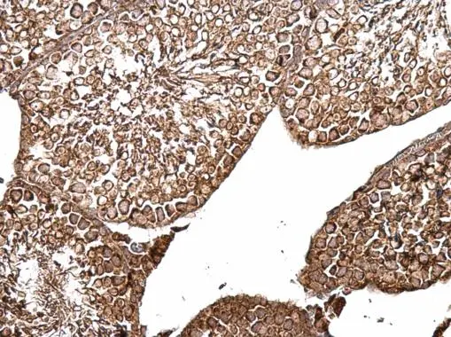
B-Raf antibody [N2C1], Internal detects B-Raf protein at cytosol on mouse testis by immunohistochemical analysis. Sample: Paraffin-embedded mouse testis. B-Raf antibody [N2C1], Internal (GTX100913) dilution: 1:500.
Antigen Retrieval: Trilogy? (EDTA based, pH 8.0) buffer, 15min
B-Raf antibody [N2C1], Internal
GTX100913
ApplicationsImmunoPrecipitation, Western Blot, ImmunoHistoChemistry, ImmunoHistoChemistry Paraffin
Product group Antibodies
ReactivityHuman, Mouse, Rat
TargetBRAF
Overview
- SupplierGeneTex
- Product NameB-Raf antibody [N2C1], Internal
- Delivery Days Customer9
- Application Supplier NoteWB: 1:500-1:10000. IHC-P: 1:100-1:1000. IP: 1:100-1:500. *Optimal dilutions/concentrations should be determined by the researcher.Not tested in other applications.
- ApplicationsImmunoPrecipitation, Western Blot, ImmunoHistoChemistry, ImmunoHistoChemistry Paraffin
- CertificationResearch Use Only
- ClonalityPolyclonal
- Concentration0.21 mg/ml
- ConjugateUnconjugated
- Gene ID673
- Target nameBRAF
- Target descriptionB-Raf proto-oncogene, serine/threonine kinase
- Target synonymsB-RAF1, B-raf, BRAF-1, BRAF1, NS7, RAFB1, serine/threonine-protein kinase B-raf, 94 kDa B-raf protein, B-Raf proto-oncogene serine/threonine-protein kinase (p94), B-Raf serine/threonine-protein, murine sarcoma viral (v-raf) oncogene homolog B1, proto-oncogene B-Raf, v-raf murine sarcoma viral oncogene homolog B, v-raf murine sarcoma viral oncogene homolog B1
- HostRabbit
- IsotypeIgG
- Protein IDP15056
- Protein NameSerine/threonine-protein kinase B-raf
- Scientific DescriptionThis gene encodes a protein belonging to the raf/mil family of serine/threonine protein kinases. This protein plays a role in regulating the MAP kinase/ERKs signaling pathway, which affects cell division, differentiation, and secretion. Mutations in this gene are associated with cardiofaciocutaneous syndrome, a disease characterized by heart defects, mental retardation and a distinctive facial appearance. Mutations in this gene have also been associated with various cancers, including non-Hodgkin lymphoma, colorectal cancer, malignant melanoma, thyroid carcinoma, non-small cell lung carcinoma, and adenocarcinoma of lung. A pseudogene, which is located on chromosome X, has been identified for this gene. [provided by RefSeq]
- ReactivityHuman, Mouse, Rat
- Storage Instruction-20°C or -80°C,2°C to 8°C
- UNSPSC41116161
Datasheet
Related products
Product group Antibodies
Anti-B-RAF AntibodyA95925
ApplicationsWestern Blot, ELISA, ImmunoHistoChemistry
ReactivityHuman, Mouse, Rat
- SizePrice
Product group Antibodies

B-RAF (Phospho-Thr599) AntibodyABX012468
ApplicationsELISA, ImmunoHistoChemistry
- SizePrice
Product group Antibodies

Anti-BRAF V600E protein [600E-7]AB03009-1.1-BT
ApplicationsELISA
ReactivityHuman
TargetBRAF
- SizePrice
Product group Antibodies
Anti-BRAF Antibody144-60889
ApplicationsImmunoFluorescence, Western Blot, ImmunoHistoChemistry
ReactivityHuman, Mouse, Rat
TargetBRAF
- SizePrice

Product group Antibodies
Anti-BRAF AntibodyAMAB91257
ApplicationsWestern Blot, ImmunoHistoChemistry
ReactivityHuman
TargetBRAF
- SizePrice
Product group Antibodies
ApplicationsImmunoFluorescence, ImmunoHistoChemistry, ImmunoHistoChemistry Frozen, ImmunoHistoChemistry Paraffin
TargetBRAF
- SizePrice
Product group Antibodies
BRAF AntibodyCSB-PA002791LA01HU
ApplicationsImmunoFluorescence, ELISA, ImmunoHistoChemistry
ReactivityHuman
TargetBRAF
- SizePrice
Product group Antibodies
Braf Polyclonal AntibodyCAC08261
ApplicationsImmunoFluorescence, ELISA, ImmunoHistoChemistry
TargetBRAF
- SizePrice
Product group Antibodies
B-Raf antibodyGTX134824
ApplicationsWestern Blot, ImmunoHistoChemistry, ImmunoHistoChemistry Paraffin
ReactivityHuman
TargetBRAF
- SizePrice