

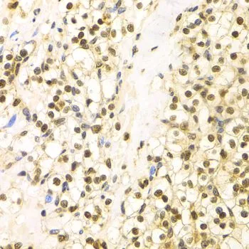
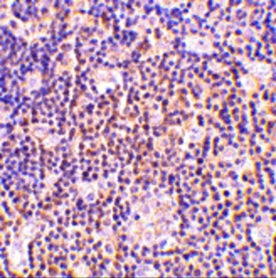
IHC-P analysis of human kidney cancer tissue using GTX30082 Bag1 antibody. Dilution : 1:100


IHC-P analysis of human kidney cancer tissue using GTX30082 Bag1 antibody. Dilution : 1:100
Bag1 antibody
GTX30082
ApplicationsImmunoFluorescence, Western Blot, ImmunoCytoChemistry, ImmunoHistoChemistry, ImmunoHistoChemistry Paraffin
Product group Antibodies
ReactivityHuman, Mouse
TargetBAG1
Overview
- SupplierGeneTex
- Product NameBag1 antibody
- Delivery Days Customer7
- Application Supplier NoteWB: 1:500 - 1:2000. ICC/IF: 1:20 - 1:100. IHC-P: 1:50 - 1:200. *Optimal dilutions/concentrations should be determined by the researcher.Not tested in other applications.
- ApplicationsImmunoFluorescence, Western Blot, ImmunoCytoChemistry, ImmunoHistoChemistry, ImmunoHistoChemistry Paraffin
- CertificationResearch Use Only
- ClonalityPolyclonal
- ConjugateUnconjugated
- Gene ID573
- Target nameBAG1
- Target descriptionBAG cochaperone 1
- Target synonymsBAG-1, HAP, RAP46, BAG family molecular chaperone regulator 1, BCL2 associated athanogene 1, BCL2-associated athanogene, Bcl-2 associating athanogene-1 protein, Bcl-2-binding protein, glucocortoid receptor-associated protein RAP46, receptor-associated protein, 46-KD
- HostRabbit
- IsotypeIgG
- Protein IDQ99933
- Protein NameBAG family molecular chaperone regulator 1
- Scientific DescriptionThe oncogene BCL2 is a membrane protein that blocks a step in a pathway leading to apoptosis or programmed cell death. The protein encoded by this gene binds to BCL2 and is referred to as BCL2-associated athanogene. It enhances the anti-apoptotic effects of BCL2 and represents a link between growth factor receptors and anti-apoptotic mechanisms. Multiple protein isoforms are encoded by this mRNA through the use of a non-AUG (CUG) initiation codon, and three alternative downstream AUG initiation codons. A related pseudogene has been defined on chromosome X. [provided by RefSeq, Feb 2010]
- ReactivityHuman, Mouse
- Storage Instruction-20°C or -80°C,2°C to 8°C
- UNSPSC41116161
References
- An increase in BAG-1 by PD-L1 confers resistance to tyrosine kinase inhibitor in non-small cell lung cancer via persistent activation of ERK signalling. Lin PL et al., 2017 Nov, Eur J CancerRead this paper
Datasheet
Related products
Product group Antibodies
Anti-BAG1 AntibodyA101545
ApplicationsWestern Blot, ELISA
ReactivityHuman
- SizePrice

Product group Antibodies
Anti-Bag1 Antibody Picoband(r)A02423-2-CARRIER-FREE
ApplicationsWestern Blot, ImmunoHistoChemistry
ReactivityHuman, Mouse, Rat
TargetBAG1
- SizePrice
Product group Antibodies
Anti-BAG1 Antibody144-01104
ApplicationsImmunoFluorescence, Western Blot, ImmunoHistoChemistry
ReactivityHuman, Mouse
TargetBAG1
- SizePrice
Product group Antibodies
ApplicationsFlow Cytometry, Western Blot, ImmunoCytoChemistry
ReactivityHuman, Mouse, Rat
TargetBAG1
- SizePrice
Product group Antibodies
BAG1 AntibodyCSB-PA005575
ApplicationsWestern Blot, ELISA
ReactivityHuman
TargetBAG1
- SizePrice
Product group Antibodies

BAG1 / BAG-1 AntibodyLS-C403776
ApplicationsWestern Blot, ELISA, ImmunoHistoChemistry
ReactivityHuman
TargetBAG1
- SizePrice
Product group Antibodies
Bag1 antibodyGTX31736
ApplicationsWestern Blot, ELISA, ImmunoHistoChemistry, ImmunoHistoChemistry Paraffin
ReactivityHuman, Mouse, Rat
TargetBAG1
- SizePrice
Product group Antibodies
Bag1 antibodyGTX31737
ApplicationsWestern Blot, ELISA, ImmunoHistoChemistry, ImmunoHistoChemistry Paraffin
ReactivityHuman, Mouse, Rat
TargetBAG1
- SizePrice
Product group Antibodies
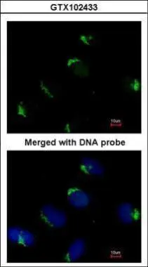
Bag1 antibody [N3C3]GTX102433
ApplicationsImmunoFluorescence, Western Blot, ImmunoCytoChemistry
ReactivityHuman
TargetBAG1
- SizePrice
Product group Antibodies
Bag1 antibody [N2C3]GTX112873
ApplicationsWestern Blot, ImmunoHistoChemistry, ImmunoHistoChemistry Paraffin
ReactivityHuman
TargetBAG1
- SizePrice