

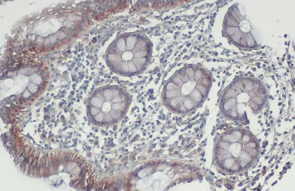
BAK antibody [N1N2], N-term detects BAK protein at mitochondria by immunohistochemical analysis. Sample: Paraffin-embedded human colon. BAK stained by BAK antibody [N1N2], N-term (GTX100063) diluted at 1:500. Antigen Retrieval: Citrate buffer, pH 6.0, 15 min
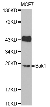
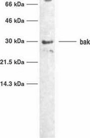
![Various whole cell extracts (30 μg) were separated by 12% SDS-PAGE, and the membrane was blotted with BAK antibody [N1N2], N-term (GTX100063) diluted at 1:1000. The HRP-conjugated anti-rabbit IgG antibody (GTX213110-01) was used to detect the primary antibody, and the signal was developed with Trident ECL plus-Enhanced.](https://www.genetex.com/upload/website/prouct_img/normal/GTX100063/GTX100063_39568_20210319_WB_w_23053123_211.webp "Various whole cell extracts (30 μg) were separated by 12% SDS-PAGE, and the membrane was blotted with BAK antibody [N1N2], N-term (GTX100063) diluted at 1:1000. The HRP-conjugated anti-rabbit IgG antibody (GTX213110-01) was used to detect the primary antibody, and the signal was developed with Trident ECL plus-Enhanced.")
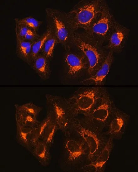
 antibody(10 μg/ml).
Antigen Retrieval: Trilogy? (EDTA based, pH 8.0) buffer, 15min")
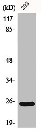
![Wild-type (WT) and BAK knockout (KO) CHO cell extracts (30 μg) were separated by 12% SDS-PAGE, and the membrane was blotted with BAK antibody [N1N2], N-term (GTX100063) diluted at 1:1000. The HRP-conjugated anti-rabbit IgG antibody (GTX213110-01) was used to detect the primary antibody.](https://www.genetex.com/upload/website/prouct_img/normal/GTX100063/GTX100063_39568_20250822_WB_KO_watermark_25082900_318.webp "Wild-type (WT) and BAK knockout (KO) CHO cell extracts (30 μg) were separated by 12% SDS-PAGE, and the membrane was blotted with BAK antibody [N1N2], N-term (GTX100063) diluted at 1:1000. The HRP-conjugated anti-rabbit IgG antibody (GTX213110-01) was used to detect the primary antibody.")
BAK antibody [N1N2], N-term detects BAK protein at mitochondria by immunohistochemical analysis. Sample: Paraffin-embedded human colon. BAK stained by BAK antibody [N1N2], N-term (GTX100063) diluted at 1:500. Antigen Retrieval: Citrate buffer, pH 6.0, 15 min
BAK antibody [N1N2], N-term
GTX100063
ApplicationsWestern Blot, ImmunoHistoChemistry, ImmunoHistoChemistry Paraffin
Product group Antibodies
ReactivityHuman
TargetBAK1
Overview
- SupplierGeneTex
- Product NameBAK antibody [N1N2], N-term
- Delivery Days Customer9
- Application Supplier NoteWB: 1:500-1:3000. IHC-P: 1:100-1:1000. *Optimal dilutions/concentrations should be determined by the researcher.Not tested in other applications.
- ApplicationsWestern Blot, ImmunoHistoChemistry, ImmunoHistoChemistry Paraffin
- CertificationResearch Use Only
- ClonalityPolyclonal
- Concentration1 mg/ml
- ConjugateUnconjugated
- Gene ID578
- Target nameBAK1
- Target descriptionBCL2 antagonist/killer 1
- Target synonymsBAK, BAK-LIKE, BCL2L7, CDN1, bcl-2 homologous antagonist/killer, BCL2-like 7 protein, apoptosis regulator BAK, bcl-2-like protein 7, bcl2-L-7, pro-apoptotic protein BAK
- HostRabbit
- IsotypeIgG
- Protein IDQ16611
- Protein NameBcl-2 homologous antagonist/killer
- Scientific DescriptionThe protein encoded by this gene belongs to the BCL2 protein family. BCL2 family members form oligomers or heterodimers and act as anti- or pro-apoptotic regulators that are involved in a wide variety of cellular activities. This protein localizes to mitochondria, and functions to induce apoptosis. It interacts with and accelerates the opening of the mitochondrial voltage-dependent anion channel, which leads to a loss in membrane potential and the release of cytochrome c. This protein also interacts with the tumor suppressor P53 after exposure to cell stress. [provided by RefSeq]
- ReactivityHuman
- Storage Instruction-20°C or -80°C,2°C to 8°C
- UNSPSC41116161
Datasheet
Related products
Product group Antibodies
Anti-BAK1 Antibody144-00204
ApplicationsImmunoFluorescence, Western Blot
ReactivityHuman, Mouse, Rat
TargetBAK1
- SizePrice
Product group Antibodies
Anti-BAK1 AntibodyA35429
ApplicationsImmunoFluorescence, Western Blot, ImmunoHistoChemistry
ReactivityHuman, Mouse, Rat
- SizePrice
Product group Antibodies
Bak Polyclonal AntibodyBS-22928R
ApplicationsWestern Blot
ReactivityHuman
TargetBAK1
- SizePrice
Product group Antibodies
BAK1 AntibodyCSB-PA000971
ApplicationsImmunoFluorescence, Western Blot, ELISA, ImmunoHistoChemistry
ReactivityHuman, Mouse
TargetBAK1
- SizePrice
Product group Antibodies

Goat anti-BAK1EB05329
ApplicationsWestern Blot, ELISA
ReactivityHuman, Mouse, Porcine, Rat
TargetBAK1
- SizePrice
Product group Antibodies
Bak1 Recombinant AntibodyCAC12015
ApplicationsFlow Cytometry, ImmunoPrecipitation, Western Blot, ELISA, ImmunoHistoChemistry
TargetBAK1
- SizePrice

Product group Antibodies
BAK antibodyGTX22273
ApplicationsWestern Blot
ReactivityHuman
TargetBAK1
- SizePrice
Product group Antibodies
BAK antibody [TC100]GTX10808
ApplicationsWestern Blot, ImmunoHistoChemistry, ImmunoHistoChemistry Frozen
ReactivityHuman
TargetBAK1
- SizePrice
Product group Antibodies
BAK antibodyGTX32463
ApplicationsImmunoFluorescence, ImmunoPrecipitation, Western Blot, ImmunoCytoChemistry
ReactivityHuman, Mouse, Rat
TargetBAK1
- SizePrice