

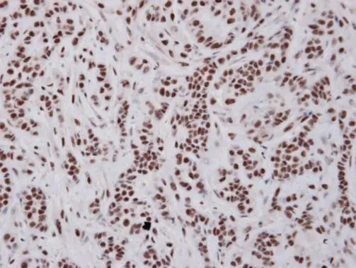
Immunohistochemical analysis of paraffin-embedded A549 xenograft , using UAP56(GTX101475) antibody at 1:100 dilution.
Antigen Retrieval: Citrate buffer, pH 6.0, 15 min
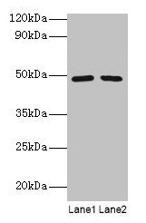
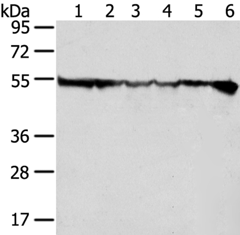
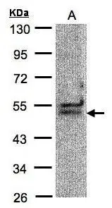
 were separated by 10% SDS-PAGE, and the membrane was blotted with BAT1 antibody (GTX101475) diluted by 1:500. The HRP-conjugated anti-rabbit IgG antibody (GTX213110-01) was used to detect the primary antibody.")
![UAP56 antibody [N1C2] detects UAP56 protein at nucleus in mouse brain by immunohistochemical analysis. Sample: Paraffin-embedded mouse brain. UAP56 antibody [N1C2] (GTX101475) diluted at 1:500.
Antigen Retrieval: Citrate buffer, pH 6.0, 15 min](https://www.genetex.com/upload/website/prouct_img/normal/GTX101475/GTX101475_40079_20150506_IHC_M_w_23060100_602.webp "UAP56 antibody [N1C2] detects UAP56 protein at nucleus in mouse brain by immunohistochemical analysis. Sample: Paraffin-embedded mouse brain. UAP56 antibody [N1C2] (GTX101475) diluted at 1:500.
Antigen Retrieval: Citrate buffer, pH 6.0, 15 min")
![BAT1 antibody [N1C2] detects BAT1 protein at nucleus by immunofluorescent analysis. Sample: HeLa cells were fixed in 4% paraformaldehyde at RT for 15 min. Green: BAT1 protein stained by BAT1 antibody [N1C2] (GTX101475) diluted at 1:500. Red: alpha Tubulin protein stained by alpha Tubulin antibody [GT114] (GTX628802) diluted at 1:1000. Scale bar = 10 μm.](https://www.genetex.com/upload/website/prouct_img/normal/GTX101475/GTX101475_42438_20160525_IFA_w_23060100_582.webp "BAT1 antibody [N1C2] detects BAT1 protein at nucleus by immunofluorescent analysis. Sample: HeLa cells were fixed in 4% paraformaldehyde at RT for 15 min. Green: BAT1 protein stained by BAT1 antibody [N1C2] (GTX101475) diluted at 1:500. Red: alpha Tubulin protein stained by alpha Tubulin antibody [GT114] (GTX628802) diluted at 1:1000. Scale bar = 10 μm.")
![BAT1 antibody [N1C2] detects BAT1 protein at nucleus in rat liver by immunohistochemical analysis. Sample: Paraffin-embedded rat liver. BAT1 antibody [N1C2] (GTX101475) diluted at 1:500.
Antigen Retrieval: Citrate buffer, pH 6.0, 15 min](https://www.genetex.com/upload/website/prouct_img/normal/GTX101475/GTX101475_42403_20160413_IHC-P_R_w_23060100_395.webp "BAT1 antibody [N1C2] detects BAT1 protein at nucleus in rat liver by immunohistochemical analysis. Sample: Paraffin-embedded rat liver. BAT1 antibody [N1C2] (GTX101475) diluted at 1:500.
Antigen Retrieval: Citrate buffer, pH 6.0, 15 min")
![UAP56 antibody [N1C2] detects UAP56 protein at nucleus in mouse ovary by immunohistochemical analysis. Sample: Paraffin-embedded mouse ovary. UAP56 antibody [N1C2] (GTX101475) diluted at 1:500.
Antigen Retrieval: Citrate buffer, pH 6.0, 15 min](https://www.genetex.com/upload/website/prouct_img/normal/GTX101475/GTX101475_40079_20150506_IHC_M_2_w_23060100_209.webp "UAP56 antibody [N1C2] detects UAP56 protein at nucleus in mouse ovary by immunohistochemical analysis. Sample: Paraffin-embedded mouse ovary. UAP56 antibody [N1C2] (GTX101475) diluted at 1:500.
Antigen Retrieval: Citrate buffer, pH 6.0, 15 min")


Immunohistochemical analysis of paraffin-embedded A549 xenograft , using UAP56(GTX101475) antibody at 1:100 dilution.
Antigen Retrieval: Citrate buffer, pH 6.0, 15 min
BAT1 antibody [N1C2]
GTX101475
ApplicationsImmunoFluorescence, Western Blot, ImmunoCytoChemistry, ImmunoHistoChemistry, ImmunoHistoChemistry Paraffin
Product group Antibodies
ReactivityHuman, Mouse, Rat
TargetDDX39B
Overview
- SupplierGeneTex
- Product NameBAT1 antibody [N1C2]
- Delivery Days Customer9
- Application Supplier NoteWB: 1:500-1:3000. ICC/IF: 1:100-1:1000. IHC-P: 1:100-1:1000. *Optimal dilutions/concentrations should be determined by the researcher.Not tested in other applications.
- ApplicationsImmunoFluorescence, Western Blot, ImmunoCytoChemistry, ImmunoHistoChemistry, ImmunoHistoChemistry Paraffin
- CertificationResearch Use Only
- ClonalityPolyclonal
- Concentration0.43 mg/ml
- ConjugateUnconjugated
- Gene ID7919
- Target nameDDX39B
- Target descriptionDExD-box helicase 39B
- Target synonymsBAT1, D6S81E, UAP56, spliceosome RNA helicase DDX39B, 56 kDa U2AF65-associated protein, ATP-dependent RNA helicase p47, DEAD (Asp-Glu-Ala-Asp) box polypeptide 39B, DEAD-box helicase 39B, HLA-B-associated transcript 1 protein, nuclear RNA helicase (DEAD family), spliceosome RNA helicase BAT1
- HostRabbit
- IsotypeIgG
- Scientific DescriptionThis gene encodes a member of the DEAD box family of RNA-dependent ATPases that mediate ATP hydrolysis during pre-mRNA splicing. The encoded protein is an essential splicing factor required for association of U2 small nuclear ribonucleoprotein with pre-mRNA, and also plays an important role in mRNA export from the nucleus to the cytoplasm. A cluster of genes, BAT1-BAT5, is localized in the vicinity of the genes for TNF alpha and TNF beta. These genes are all within the human major histocompatibility complex class III region. Mutations in this gene may be associated with rheumatoid arthritis. Alternatively spliced transcript variants encoding the same protein have been described. [provided by RefSeq]
- ReactivityHuman, Mouse, Rat
- Storage Instruction-20°C or -80°C,2°C to 8°C
- UNSPSC41116161
Datasheet
Related products
Product group Antibodies
DDX39B AntibodyCSB-PA00304A0RB
ApplicationsImmunoFluorescence, Western Blot, ELISA, ImmunoHistoChemistry
ReactivityHuman
TargetDDX39B
- SizePrice
Product group Antibodies
Anti-DDX39B AntibodyA37361
ApplicationsWestern Blot, ImmunoHistoChemistry
ReactivityHuman
- SizePrice

Product group Antibodies
ApplicationsFlow Cytometry, ImmunoFluorescence, Western Blot, ImmunoCytoChemistry, ImmunoHistoChemistry
ReactivityHuman, Mouse, Rat
TargetDDX39B
- SizePrice

Product group Antibodies
Anti-DDX39B AntibodyHPA055334
ApplicationsWestern Blot, ImmunoCytoChemistry, ImmunoHistoChemistry
ReactivityHuman
TargetDDX39B
- SizePrice
Product group Antibodies

DDX39B / UAP56 AntibodyLS-C409892
ApplicationsWestern Blot, ImmunoHistoChemistry
ReactivityHuman, Mouse, Rat
TargetDDX39B
- SizePrice
Product group Antibodies
Ddx39B Polyclonal AntibodyCAC08265
ApplicationsImmunoFluorescence, Western Blot, ELISA, ImmunoHistoChemistry
TargetDDX39B
- SizePrice
![WB analysis of HEK293T cells transfected with BAT1 plasmid (Right) or empty vector (Left) for 48 hrs using GTX84837 BAT1 antibody [2C5]. Loading : 5 ug per lane](https://www.genetex.com/upload/website/prouct_img/normal/GTX84837/GTX84837_4656_WB_w_23061420_276.webp)
Product group Antibodies
BAT1 antibody [2C5]GTX84837
ApplicationsWestern Blot, ImmunoHistoChemistry, ImmunoHistoChemistry Paraffin
ReactivityCanine, Human, Monkey, Mouse
TargetDDX39B
- SizePrice
Product group Antibodies
BAT1 antibody [C1C3]GTX101476
ApplicationsWestern Blot
ReactivityHuman
TargetDDX39B
- SizePrice