

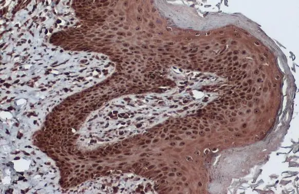
Bcl-2 antibody detects Bcl-2 protein at cytoplasm and nucleus by immunohistochemical analysis. Sample: Paraffin-embedded dog skin. Bcl-2 stained by Bcl-2 antibody (GTX100064) diluted at 1:500. Antigen Retrieval: Citrate buffer, pH 6.0, 15 min
 was separated by 12% SDS-PAGE, and the membrane was blotted with Bcl-2 antibody (GTX100064) diluted at 1:1000. The HRP-conjugated anti-rabbit IgG antibody (GTX213110-01) was used to detect the primary antibody, and the signal was developed with Trident ECL plus-Enhanced.")
 was separated by 12% SDS-PAGE, and the membrane was blotted with Bcl-2 antibody (GTX100064) diluted at 1:1000. The HRP-conjugated anti-rabbit IgG antibody (GTX213110-01) was used to detect the primary antibody, and the signal was developed with Trident ECL plus-Enhanced.")
 diluted at 1:500. Antigen Retrieval: Citrate buffer, pH 6.0, 15 min")
![Whole cell extract (30 μg) was separated by 12% SDS-PAGE, and the membrane was blotted with Bcl-2 antibody [N1N2], N-term (GTX100064) diluted at 1:1000. The HRP-conjugated anti-rabbit IgG antibody (GTX213110-01) was used to detect the primary antibody.](https://www.genetex.com/upload/website/prouct_img/normal/GTX100064/GTX100064_43012_20210806_WB_M_w_23053123_854.webp "Whole cell extract (30 μg) was separated by 12% SDS-PAGE, and the membrane was blotted with Bcl-2 antibody [N1N2], N-term (GTX100064) diluted at 1:1000. The HRP-conjugated anti-rabbit IgG antibody (GTX213110-01) was used to detect the primary antibody.")
![Bcl-2 antibody [N1N2], N-term detects Bcl-2 protein at cytoplasm and nucleus by immunofluorescent analysis. Sample: THP-1 cells were fixed in 4% paraformaldehyde at RT for 15 min. Green: Bcl-2 stained by Bcl-2 antibody [N1N2], N-term (GTX100064) diluted at 1:500. Blue: Fluoroshield with DAPI (GTX30920).](https://www.genetex.com/upload/website/prouct_img/normal/GTX100064/GTX100064_42915_20180919_ICC_IF_w_23053123_598.webp "Bcl-2 antibody [N1N2], N-term detects Bcl-2 protein at cytoplasm and nucleus by immunofluorescent analysis. Sample: THP-1 cells were fixed in 4% paraformaldehyde at RT for 15 min. Green: Bcl-2 stained by Bcl-2 antibody [N1N2], N-term (GTX100064) diluted at 1:500. Blue: Fluoroshield with DAPI (GTX30920).")
![Various whole cell extracts (30 μg) were separated by 12% SDS-PAGE, and the membrane was blotted with Bcl-2 antibody [N1N2], N-term (GTX100064) diluted at 1:500. The HRP-conjugated anti-rabbit IgG antibody (GTX213110-01) was used to detect the primary antibody.](https://www.genetex.com/upload/website/prouct_img/normal/GTX100064/GTX100064_42970_20200403_WB_competitor_watermark_w_23053123_457.webp "Various whole cell extracts (30 μg) were separated by 12% SDS-PAGE, and the membrane was blotted with Bcl-2 antibody [N1N2], N-term (GTX100064) diluted at 1:500. The HRP-conjugated anti-rabbit IgG antibody (GTX213110-01) was used to detect the primary antibody.")
![Non-transfected (–) and transfected (+) 293T whole cell extracts (30 μg) were separated by 12% SDS-PAGE, and the membrane was blotted with Bcl-2 antibody [N1N2], N-term (GTX100064) diluted at 1:500. The HRP-conjugated anti-rabbit IgG antibody (GTX213110-01) was used to detect the primary antibody.](https://www.genetex.com/upload/website/prouct_img/normal/GTX100064/GTX100064_42915_20181126_WB_B_w_23053123_496.webp "Non-transfected (–) and transfected (+) 293T whole cell extracts (30 μg) were separated by 12% SDS-PAGE, and the membrane was blotted with Bcl-2 antibody [N1N2], N-term (GTX100064) diluted at 1:500. The HRP-conjugated anti-rabbit IgG antibody (GTX213110-01) was used to detect the primary antibody.")
![Rat tissue extract (50 μg) was separated by 12% SDS-PAGE, and the membrane was blotted with Bcl-2 antibody [N1N2], N-term (GTX100064) diluted at 1:1000. The HRP-conjugated anti-rabbit IgG antibody (GTX213110-01) was used to detect the primary antibody, and the signal was developed with Trident ECL plus-Enhanced.](https://www.genetex.com/upload/website/prouct_img/normal/GTX100064/GTX100064_43012_20210806_WB_R_pancrease_w_23053123_738.webp "Rat tissue extract (50 μg) was separated by 12% SDS-PAGE, and the membrane was blotted with Bcl-2 antibody [N1N2], N-term (GTX100064) diluted at 1:1000. The HRP-conjugated anti-rabbit IgG antibody (GTX213110-01) was used to detect the primary antibody, and the signal was developed with Trident ECL plus-Enhanced.")
![Various whole cell extracts (30 μg) were separated by 12% SDS-PAGE, and the membrane was blotted with Bcl-2 antibody [N1N2], N-term (GTX100064) diluted at 1:500. The HRP-conjugated anti-rabbit IgG antibody (GTX213110-01) was used to detect the primary antibody. Corresponding RNA expression data for the same cell lines are based on Human Protein Atlas program.](https://www.genetex.com/upload/website/prouct_img/normal/GTX100064/GTX100064_43012_20190830_WB_TPM_watermark_w_23053123_528.webp "Various whole cell extracts (30 μg) were separated by 12% SDS-PAGE, and the membrane was blotted with Bcl-2 antibody [N1N2], N-term (GTX100064) diluted at 1:500. The HRP-conjugated anti-rabbit IgG antibody (GTX213110-01) was used to detect the primary antibody. Corresponding RNA expression data for the same cell lines are based on Human Protein Atlas program.")
Bcl-2 antibody detects Bcl-2 protein at cytoplasm and nucleus by immunohistochemical analysis. Sample: Paraffin-embedded dog skin. Bcl-2 stained by Bcl-2 antibody (GTX100064) diluted at 1:500. Antigen Retrieval: Citrate buffer, pH 6.0, 15 min
Bcl-2 antibody [N1N2], N-term
GTX100064
ApplicationsImmunoFluorescence, Western Blot, ImmunoCytoChemistry, ImmunoHistoChemistry, ImmunoHistoChemistry Paraffin
Product group Antibodies
ReactivityCanine, Feline, Human, Mouse, Rat
TargetBCL2
Overview
- SupplierGeneTex
- Product NameBcl-2 antibody [N1N2], N-term
- Delivery Days Customer9
- Application Supplier NoteWB: 1:500-1:3000. ICC/IF: 1:100-1:1000. IHC-P: 1:100-1:1000. *Optimal dilutions/concentrations should be determined by the researcher.Not tested in other applications.
- ApplicationsImmunoFluorescence, Western Blot, ImmunoCytoChemistry, ImmunoHistoChemistry, ImmunoHistoChemistry Paraffin
- CertificationResearch Use Only
- ClonalityPolyclonal
- Concentration0.2 mg/ml
- ConjugateUnconjugated
- Gene ID596
- Target nameBCL2
- Target descriptionBCL2 apoptosis regulator
- Target synonymsBcl-2, PPP1R50, apoptosis regulator Bcl-2, B-cell CLL/lymphoma 2, protein phosphatase 1, regulatory subunit 50
- HostRabbit
- IsotypeIgG
- Protein IDP10415
- Protein NameApoptosis regulator Bcl-2
- Scientific DescriptionThis gene encodes an integral outer mitochondrial membrane protein that blocks the apoptotic death of some cells such as lymphocytes. Constitutive expression of BCL2, such as in the case of translocation of BCL2 to Ig heavy chain locus, is thought to be the cause of follicular lymphoma. Two transcript variants, produced by alternate splicing, differ in their C-terminal ends. [provided by RefSeq]
- ReactivityCanine, Feline, Human, Mouse, Rat
- Storage Instruction-20°C or -80°C,2°C to 8°C
- UNSPSC41116161
Datasheet
Related products

Product group Antibodies
Anti-Bcl-2/BCL2 Antibody Picoband(r)A00040-2-CARRIER-FREE
ApplicationsFlow Cytometry, ImmunoFluorescence, Western Blot, ELISA, ImmunoCytoChemistry
ReactivityHuman
TargetBCL2
- SizePrice

Product group Antibodies
Anti-BCL2 AntibodyAMAB91492
ApplicationsWestern Blot, ImmunoHistoChemistry
ReactivityHuman
TargetBCL2
- SizePrice
Product group Antibodies

Anti-BCL2 [bcl-2/100]Ab01321-1.1
ApplicationsImmunoPrecipitation, Western Blot, ImmunoHistoChemistry
ReactivityHuman
TargetBCL2
- SizePrice
Product group Antibodies
ApplicationsImmunoPrecipitation, Western Blot, ImmunoHistoChemistry
ReactivityHuman, Mouse, Rat
- SizePrice
Product group Antibodies

ApplicationsWestern Blot, ELISA
ReactivityHuman
TargetBCL2
- SizePrice
Product group Antibodies

Goat anti-BCL2EB06444
ApplicationsWestern Blot, ELISA
ReactivityCanine, Human, Mouse, Rat
TargetBCL2
- SizePrice
Product group Antibodies
BCL2 Monoclonal AntibodyCSB-MA000196
ApplicationsWestern Blot, ELISA, ImmunoHistoChemistry
ReactivityHuman, Mouse, Rat
TargetBCL2
- SizePrice
Product group Antibodies
Bcl-2 antibodyGTX27973
ApplicationsImmunoPrecipitation, Western Blot, ImmunoHistoChemistry
ReactivityHuman, Mouse, Rat
TargetBCL2
- SizePrice
![WB analysis of Jurkat cell lysate using GTX31237 Bcl-2 antibody [100].](https://www.genetex.com/upload/website/prouct_img/normal/GTX31237/GTX31237_138_WB_w_23060722_726.webp)
Product group Antibodies
Bcl-2 antibody [100]GTX31237
ApplicationsFlow Cytometry, ImmunoPrecipitation, Western Blot, ImmunoHistoChemistry, ImmunoHistoChemistry Frozen, ImmunoHistoChemistry Paraffin
ReactivityHuman
TargetBCL2
- SizePrice