

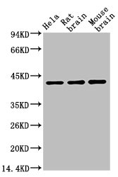
Various whole cell extracts (30 μg) were separated by 10% SDS-PAGE, and the membrane was blotted with beta Actin antibody [HL1926] (GTX637675) diluted at 1:50000. The HRP-conjugated anti-rabbit IgG antibody (GTX213110-01) was used to detect the primary antibody.
![Whole cell extract (30 μg) was separated by 10% SDS-PAGE, and the membrane was blotted with beta Actin antibody [HL1926] (GTX637675) diluted at 1:10000. The HRP-conjugated anti-rabbit IgG antibody (GTX213110-01) was used to detect the primary antibody.](https://www.genetex.com/upload/website/prouct_img/normal/GTX637675/GTX637675_T-44837_20221111_WB_Drosophila_22111518_843.webp "Whole cell extract (30 μg) was separated by 10% SDS-PAGE, and the membrane was blotted with beta Actin antibody [HL1926] (GTX637675) diluted at 1:10000. The HRP-conjugated anti-rabbit IgG antibody (GTX213110-01) was used to detect the primary antibody.")
![Various whole cell extracts (30 μg) were separated by 10% SDS-PAGE, and the membrane was blotted with beta Actin antibody [HL1926] (GTX637675) diluted at 1:10000. The HRP-conjugated anti-rabbit IgG antibody (GTX213110-01) was used to detect the primary antibody.](https://www.genetex.com/upload/website/prouct_img/normal/GTX637675/GTX637675_T-44837_20221111_WB_C_D_22111518_626.webp "Various whole cell extracts (30 μg) were separated by 10% SDS-PAGE, and the membrane was blotted with beta Actin antibody [HL1926] (GTX637675) diluted at 1:10000. The HRP-conjugated anti-rabbit IgG antibody (GTX213110-01) was used to detect the primary antibody.")
![beta Actin antibody [HL1926] detects beta Actin protein at cell membrane by immunohistochemical analysis. Sample: Paraffin-embedded mouse intestine. beta Actin stained by beta Actin antibody [HL1926] (GTX637675) diluted at 1:100. Antigen Retrieval: Citrate buffer, pH 6.0, 15 min](https://www.genetex.com/upload/website/prouct_img/normal/GTX637675/GTX637675_T-44837_20221111_IHC-P_M_22111518_644.webp "beta Actin antibody [HL1926] detects beta Actin protein at cell membrane by immunohistochemical analysis. Sample: Paraffin-embedded mouse intestine. beta Actin stained by beta Actin antibody [HL1926] (GTX637675) diluted at 1:100. Antigen Retrieval: Citrate buffer, pH 6.0, 15 min")
![Whole cell extract (30 μg) was separated by 10% SDS-PAGE, and the membrane was blotted with beta Actin antibody [HL1926] (GTX637675) diluted at 1:10000. The HRP-conjugated anti-rabbit IgG antibody (GTX213110-01) was used to detect the primary antibody.](https://www.genetex.com/upload/website/prouct_img/normal/GTX637675/GTX637675_T-44837_20221111_WB_Z_22111518_555.webp "Whole cell extract (30 μg) was separated by 10% SDS-PAGE, and the membrane was blotted with beta Actin antibody [HL1926] (GTX637675) diluted at 1:10000. The HRP-conjugated anti-rabbit IgG antibody (GTX213110-01) was used to detect the primary antibody.")
![beta Actin antibody [HL1926] detects beta Actin protein by immunofluorescent analysis. Sample: HeLa cells were fixed in ice-cold MeOH for 10 min. Green: beta Actin stained by beta Actin antibody [HL1926] (GTX637675) diluted at 1:500. Blue: Fluoroshield with DAPI (GTX30920).](https://www.genetex.com/upload/website/prouct_img/normal/GTX637675/GTX637675_44900_20221230_ICC_IF_22122901_487.webp "beta Actin antibody [HL1926] detects beta Actin protein by immunofluorescent analysis. Sample: HeLa cells were fixed in ice-cold MeOH for 10 min. Green: beta Actin stained by beta Actin antibody [HL1926] (GTX637675) diluted at 1:500. Blue: Fluoroshield with DAPI (GTX30920).")
![Various whole cell extracts (30 μg) were separated by 10% SDS-PAGE, and the membrane was blotted with beta Actin antibody [HL1926] (GTX637675) diluted at 1:50000. The HRP-conjugated anti-rabbit IgG antibody (GTX213110-01) was used to detect the primary antibody.](https://www.genetex.com/upload/website/prouct_img/normal/GTX637675/GTX637675_44900_20230915_WB_23091901_695.webp "Various whole cell extracts (30 μg) were separated by 10% SDS-PAGE, and the membrane was blotted with beta Actin antibody [HL1926] (GTX637675) diluted at 1:50000. The HRP-conjugated anti-rabbit IgG antibody (GTX213110-01) was used to detect the primary antibody.")
![Various whole cell extracts (30 μg) were separated by 10% SDS-PAGE, and the membranes were blotted with beta Actin antibody [HL1926] (GTX637675) diluted at 1:10000. The HRP-conjugated anti-rabbit IgG antibody (GTX213110-01) was used to detect the primary antibody.](https://www.genetex.com/upload/website/prouct_img/normal/GTX637675/GTX637675_45180_20231110_WB_multiple_species_23112021_692.webp "Various whole cell extracts (30 μg) were separated by 10% SDS-PAGE, and the membranes were blotted with beta Actin antibody [HL1926] (GTX637675) diluted at 1:10000. The HRP-conjugated anti-rabbit IgG antibody (GTX213110-01) was used to detect the primary antibody.")
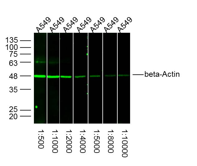
![Various whole cell extracts were separated by 10% SDS-PAGE, and the membrane was blotted with beta Actin antibody [HL1926] (GTX637675) diluted at 1:100000. The HRP-conjugated anti-rabbit IgG antibody (GTX213110-01) was used to detect the primary antibody.](https://www.genetex.com/upload/website/prouct_img/normal/GTX637675/GTX637675_45467_20240712_WB_Sensitivity_25092520_257.webp "Various whole cell extracts were separated by 10% SDS-PAGE, and the membrane was blotted with beta Actin antibody [HL1926] (GTX637675) diluted at 1:100000. The HRP-conjugated anti-rabbit IgG antibody (GTX213110-01) was used to detect the primary antibody.")
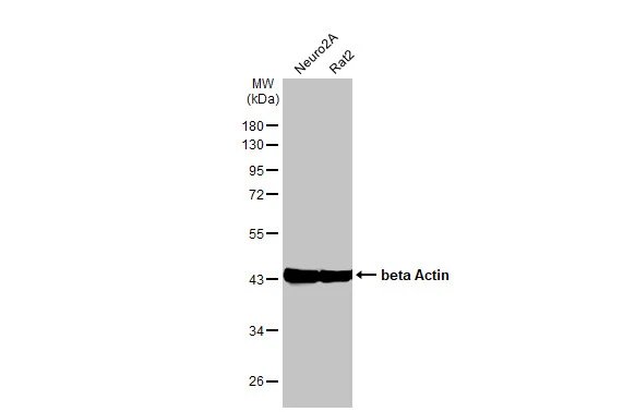
Various whole cell extracts (30 μg) were separated by 10% SDS-PAGE, and the membrane was blotted with beta Actin antibody [HL1926] (GTX637675) diluted at 1:50000. The HRP-conjugated anti-rabbit IgG antibody (GTX213110-01) was used to detect the primary antibody.
beta Actin antibody [HL1926]
GTX637675
ApplicationsImmunoFluorescence, Western Blot, ImmunoCytoChemistry, ImmunoHistoChemistry, ImmunoHistoChemistry Paraffin
Product group Antibodies
ReactivityCanine, Drosophila, Feline, Human, Monkey, Mouse, Rabbit, Rat, Zebra Fish
TargetACTB
Overview
- SupplierGeneTex
- Product Namebeta Actin antibody [HL1926]
- Delivery Days Customer9
- Application Supplier NoteWB: 1:5000-1:50000. *Optimal dilutions/concentrations should be determined by the researcher.Not tested in other applications.
- ApplicationsImmunoFluorescence, Western Blot, ImmunoCytoChemistry, ImmunoHistoChemistry, ImmunoHistoChemistry Paraffin
- CertificationResearch Use Only
- ClonalityMonoclonal
- Clone IDHL1926
- Concentration1 mg/ml
- ConjugateUnconjugated
- Gene ID60
- Target nameACTB
- Target descriptionactin beta
- Target synonymsBKRNS, BNS, BRWS1, CSMH, DDS1, PS1TP5BP1, THC8, actin, cytoplasmic 1, I(2)-actin, PS1TP5-binding protein 1, beta cytoskeletal actin
- HostRabbit
- IsotypeIgG
- Protein IDP60709
- Protein NameActin, cytoplasmic 1
- Scientific DescriptionThis gene encodes one of six different actin proteins. Actins are highly conserved proteins that are involved in cell motility, structure, integrity, and intercellular signaling. The encoded protein is a major constituent of the contractile apparatus and one of the two nonmuscle cytoskeletal actins that are ubiquitously expressed. Mutations in this gene cause Baraitser-Winter syndrome 1, which is characterized by intellectual disability with a distinctive facial appearance in human patients. Numerous pseudogenes of this gene have been identified throughout the human genome. [provided by RefSeq, Aug 2017]
- ReactivityCanine, Drosophila, Feline, Human, Monkey, Mouse, Rabbit, Rat, Zebra Fish
- Storage Instruction-20°C or -80°C,2°C to 8°C
- UNSPSC41116161
Datasheet
Related products
Product group Antibodies

Anti-Beta-actin [Fab 19, actin]Ab02221-1.1
ApplicationsImmunoFluorescence, ELISA
ReactivityHuman
TargetACTB
- SizePrice
Product group Antibodies

Actin-pan AntibodyABX013019
ApplicationsWestern Blot, ELISA, ImmunoHistoChemistry
- SizePrice
Product group Antibodies
Anti-beta Actin Antibody102-26634
ApplicationsWestern Blot
TargetACTB
- SizePrice
Product group Antibodies
Anti-beta Actin AntibodyA121645
ApplicationsImmunoFluorescence, Western Blot
ReactivityCanine, Human, Monkey, Mouse, Rat
- SizePrice

Product group Antibodies
Anti-ACTB AntibodyAMAB91241
ApplicationsWestern Blot, ImmunoCytoChemistry, ImmunoHistoChemistry
ReactivityHuman, Mouse, Rat
TargetACTB
- SizePrice
Product group Antibodies

ACTB / Beta Actin AntibodyLS-C831486
ApplicationsWestern Blot, ImmunoHistoChemistry
ReactivityHuman, Mouse, Rat, Zebra Fish
TargetACTB
- SizePrice
Product group Antibodies
References
beta-Actin Polyclonal AntibodyBS-0061R
ApplicationsFlow Cytometry, ImmunoFluorescence, Western Blot, ImmunoCytoChemistry, ImmunoHistoChemistry, ImmunoHistoChemistry Frozen, ImmunoHistoChemistry Paraffin
ReactivityCanine, Chicken, Feline, Fish, Guinea Pig, Hamster, Human, Insect, Mouse, Porcine, Rabbit, Rat, Sheep
TargetACTB
- SizePrice
Product group Antibodies
ACTB Monoclonal AntibodyCSB-MA000187
ApplicationsWestern Blot, ELISA
ReactivityCanine, Chicken, Hamster, Human, Monkey, Mouse, Porcine, Rabbit, Rat, Sheep
TargetACTB
- SizePrice
Product group Antibodies
Actb Polyclonal AntibodyCAC07011
ApplicationsImmunoFluorescence, Western Blot, ELISA, ImmunoHistoChemistry
TargetACTB
- SizePrice