

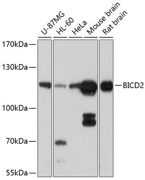
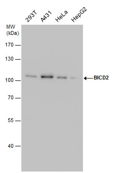
Whole cell extract (30 μg) was separated by 7.5% SDS-PAGE, and the membrane was blotted with BICD2 antibody [GT10811] (GTX631994) diluted at 1:1000.
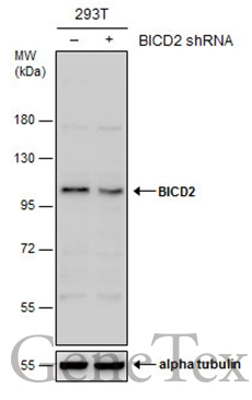
![Non-transfected (–) and transfected (+) 293T whole cell extracts (30 μg) were separated by 7.5% SDS-PAGE, and the membrane was blotted with BICD2 antibody [GT10811] (GTX631994) diluted at 1:500.](https://www.genetex.com/upload/website/prouct_img/normal/GTX631994/GTX631994_41911_20161117_WB_shRNA_watermark_w_23061202_373.webp "Non-transfected (–) and transfected (+) 293T whole cell extracts (30 μg) were separated by 7.5% SDS-PAGE, and the membrane was blotted with BICD2 antibody [GT10811] (GTX631994) diluted at 1:500.")
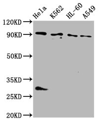
![Mouse tissue extract (50 μg) was separated by 7.5% SDS-PAGE, and the membrane was blotted with BICD2 antibody [GT10811] (GTX631994) diluted at 1:1000.](https://www.genetex.com/upload/website/prouct_img/normal/GTX631994/GTX631994_41911_20160825_WB_M_lung_w_23061202_254.webp "Mouse tissue extract (50 μg) was separated by 7.5% SDS-PAGE, and the membrane was blotted with BICD2 antibody [GT10811] (GTX631994) diluted at 1:1000.")
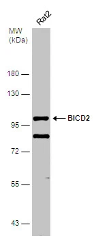
 were separated by 7.5% SDS-PAGE, and the membrane was blotted with BICD2 antibody (GTX631994) diluted by 1:1000.")
![Immunoprecipitation of BICD2 protein from A431 whole cell extracts using 5 μg of BICD2 antibody [GT10811] (GTX631994). Western blot analysis was performed using BICD2 antibody [GT10811] (GTX631994). EasyBlot anti-Mouse IgG (GTX221667-01) was used as a secondary reagent.](https://www.genetex.com/upload/website/prouct_img/normal/GTX631994/GTX631994_41911_20150710_IP_w_23061202_523.webp "Immunoprecipitation of BICD2 protein from A431 whole cell extracts using 5 μg of BICD2 antibody [GT10811] (GTX631994). Western blot analysis was performed using BICD2 antibody [GT10811] (GTX631994). EasyBlot anti-Mouse IgG (GTX221667-01) was used as a secondary reagent.")
![BICD2 antibody [GT10811] detects BICD2 protein by immunofluorescent analysis. Sample: HeLa cells were fixed in ice-cold MeOH for 5 min. Green: BICD2 stained by BICD2 antibody [GT10811] (GTX631994) diluted at 1:500. Red: alpha Tubulin, a cytoskeleton marker, stained by alpha Tubulin antibody [GT114] (GTX628802) diluted at 1:1000. Blue: Fluoroshield with DAPI (GTX30920).](https://www.genetex.com/upload/website/prouct_img/normal/GTX631994/GTX631994_41911_20240510_ICC_IF_24052202_316.webp "BICD2 antibody [GT10811] detects BICD2 protein by immunofluorescent analysis. Sample: HeLa cells were fixed in ice-cold MeOH for 5 min. Green: BICD2 stained by BICD2 antibody [GT10811] (GTX631994) diluted at 1:500. Red: alpha Tubulin, a cytoskeleton marker, stained by alpha Tubulin antibody [GT114] (GTX628802) diluted at 1:1000. Blue: Fluoroshield with DAPI (GTX30920).")
Whole cell extract (30 μg) was separated by 7.5% SDS-PAGE, and the membrane was blotted with BICD2 antibody [GT10811] (GTX631994) diluted at 1:1000.
BICD2 antibody [GT10811]
GTX631994
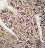
ApplicationsImmunoFluorescence, ImmunoPrecipitation, Western Blot, ImmunoCytoChemistry
Product group Antibodies
ReactivityHuman, Mouse, Rat
TargetBICD2
Overview
- SupplierGeneTex
- Product NameBICD2 antibody [GT10811]
- Delivery Days Customer9
- Application Supplier NoteWB: 1:500-1:3000. ICC/IF: 1:100-1:1000. IP: 1:100-1:500. *Optimal dilutions/concentrations should be determined by the researcher.Not tested in other applications.
- ApplicationsImmunoFluorescence, ImmunoPrecipitation, Western Blot, ImmunoCytoChemistry
- CertificationResearch Use Only
- ClonalityMonoclonal
- Clone IDGT10811
- Concentration1 mg/ml
- ConjugateUnconjugated
- Gene ID23299
- Target nameBICD2
- Target descriptionBICD cargo adaptor 2
- Target synonymsSMALED2, SMALED2A, SMALED2B, bA526D8.1, protein bicaudal D homolog 2, bic-D 2, bicaudal D homolog 2, coiled-coil protein BICD2, cytoskeleton-like bicaudal D protein homolog 2, homolog of Drosophila bicaudal D
- HostMouse
- IsotypeIgG2b
- Protein IDQ8TD16
- Protein NameProtein bicaudal D homolog 2
- Scientific DescriptionThis gene is one of two human homologs of Drosophila bicaudal-D and a member of the Bicoid family. It has been implicated in dynein-mediated, minus end-directed motility along microtubules. It has also been reported to be a phosphorylation target of NIMA related kinase 8. Two alternative splice variants have been described. [provided by RefSeq]
- ReactivityHuman, Mouse, Rat
- Storage Instruction-20°C or -80°C,2°C to 8°C
- UNSPSC41116161
Datasheet
Related products
Product group Antibodies
Anti-BICD2 AntibodyA12879
ApplicationsWestern Blot
ReactivityHuman, Mouse, Rat
- SizePrice
Product group Antibodies

BICD2 AntibodyLS-C832751
ApplicationsELISA, ImmunoHistoChemistry
ReactivityHuman
TargetBICD2
- SizePrice

Product group Antibodies
Anti-BICD2 AntibodyHPA023013
ApplicationsWestern Blot, ImmunoCytoChemistry, ImmunoHistoChemistry
ReactivityHuman, Mouse, Rat
TargetBICD2
- SizePrice

Product group Antibodies
ApplicationsWestern Blot, ELISA
ReactivityHuman, Mouse, Rat
TargetBICD2
- SizePrice
Product group Antibodies
BICD2 AntibodyCSB-PA844723LA01HU
ApplicationsImmunoFluorescence, Western Blot, ELISA, ImmunoHistoChemistry
ReactivityHuman
TargetBICD2
- SizePrice
Product group Antibodies
Bicd2 Polyclonal AntibodyCAC11464
ApplicationsImmunoFluorescence, Western Blot, ELISA, ImmunoHistoChemistry
TargetBICD2
- SizePrice
Product group Antibodies
BICD2 antibody, C-termGTX81309
ApplicationsWestern Blot, ImmunoHistoChemistry, ImmunoHistoChemistry Paraffin
ReactivityHuman
TargetBICD2
- SizePrice
Product group Antibodies
BICD2 antibodyGTX120683
ApplicationsWestern Blot, ImmunoHistoChemistry, ImmunoHistoChemistry Paraffin
ReactivityHuman, Mouse, Rat
TargetBICD2
- SizePrice
Product group Antibodies
BICD2 antibody [GT1824]GTX631995
ApplicationsImmunoPrecipitation, Western Blot
ReactivityHuman, Rat
TargetBICD2
- SizePrice