

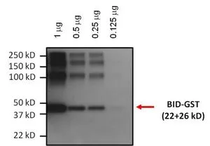
WB analysis of 0.125-1ug purified BID-GST fusion protein using GTX15667 Bid antibody [23F7]. Dilution : 1:1000
![ICC/IF analysis of HeLa cells using GTX15667 Bid antibody [23F7]. Panel e is a no primary antibody control. Green : Primary antibody Blue : Nuclei Red : Actin Fixation : 4% paraformaldehyde Permeabilization : 0.1% Trito X-100 for 10 minutes Dilution : 2 μg/ml in 0.1% BSA and incubated for 3 hours at room temperature](https://www.genetex.com/upload/website/prouct_img/normal/GTX15667/GTX15667_281_ICC-IF_w_23060620_951.webp "ICC/IF analysis of HeLa cells using GTX15667 Bid antibody [23F7]. Panel e is a no primary antibody control. Green : Primary antibody Blue : Nuclei Red : Actin Fixation : 4% paraformaldehyde Permeabilization : 0.1% Trito X-100 for 10 minutes Dilution : 2 μg/ml in 0.1% BSA and incubated for 3 hours at room temperature")
![ICC/IF analysis of Hela cells using GTX15667 Bid antibody [23F7]. Cells were probed without (left) or with(right) an antibody. Green : Primary antibody Blue : Nuclei Red : Actin Fixation : Formalin Permeabilization : 0.1% Triton X-100 in TBS for 5-10 minutes Dilution : 1:20 overnight at 4oC](https://www.genetex.com/upload/website/prouct_img/normal/GTX15667/GTX15667_283_ICC-IF_w_23060620_632.webp "ICC/IF analysis of Hela cells using GTX15667 Bid antibody [23F7]. Cells were probed without (left) or with(right) an antibody. Green : Primary antibody Blue : Nuclei Red : Actin Fixation : Formalin Permeabilization : 0.1% Triton X-100 in TBS for 5-10 minutes Dilution : 1:20 overnight at 4oC")
![ICC/IF analysis of A549 cells using GTX15667 Bid antibody [23F7]. Cells were probed without (left) or with(right) an antibody. Green : Primary antibody Blue : Nuclei Red : Actin Fixation : Formalin Permeabilization : 0.1% Triton X-100 in TBS for 5-10 minutes Dilution : 1:20 overnight at 4oC](https://www.genetex.com/upload/website/prouct_img/normal/GTX15667/GTX15667_282_ICC-IF_w_23060620_917.webp "ICC/IF analysis of A549 cells using GTX15667 Bid antibody [23F7]. Cells were probed without (left) or with(right) an antibody. Green : Primary antibody Blue : Nuclei Red : Actin Fixation : Formalin Permeabilization : 0.1% Triton X-100 in TBS for 5-10 minutes Dilution : 1:20 overnight at 4oC")
![ICC/IF analysis of NIH-3T3 cells using GTX15667 Bid antibody [23F7]. Cells were probed without (left) or with(right) an antibody. Green : Primary antibody Blue : Nuclei Fixation : Formalin Permeabilization : 0.1% Triton X-100 in TBS for 5-10 minutes Dilution : 1:20 overnight at 4oC](https://www.genetex.com/upload/website/prouct_img/normal/GTX15667/GTX15667_284_ICC-IF_w_23060620_589.webp "ICC/IF analysis of NIH-3T3 cells using GTX15667 Bid antibody [23F7]. Cells were probed without (left) or with(right) an antibody. Green : Primary antibody Blue : Nuclei Fixation : Formalin Permeabilization : 0.1% Triton X-100 in TBS for 5-10 minutes Dilution : 1:20 overnight at 4oC")
WB analysis of 0.125-1ug purified BID-GST fusion protein using GTX15667 Bid antibody [23F7]. Dilution : 1:1000
Bid antibody [23F7]
GTX15667
ApplicationsImmunoFluorescence, Western Blot, ImmunoCytoChemistry
Product group Antibodies
ReactivityHuman, Mouse
TargetBID
Overview
- SupplierGeneTex
- Product NameBid antibody [23F7]
- Delivery Days Customer9
- Application Supplier NoteWB: 1:1000. ICC/IF: 1-2 microg/ml. *Optimal dilutions/concentrations should be determined by the researcher.Not tested in other applications.
- ApplicationsImmunoFluorescence, Western Blot, ImmunoCytoChemistry
- CertificationResearch Use Only
- ClonalityMonoclonal
- Clone ID23F7
- Concentration1 mg/ml
- ConjugateUnconjugated
- Gene ID637
- Target nameBID
- Target descriptionBH3 interacting domain death agonist
- Target synonymsFP497, BH3-interacting domain death agonist, Human BID coding sequence, apoptic death agonist, desmocollin type 4, p22 BID
- HostMouse
- IsotypeIgG
- Protein IDP55957
- Protein NameBH3-interacting domain death agonist
- Scientific DescriptionThis gene encodes a death agonist that heterodimerizes with either agonist BAX or antagonist BCL2. The encoded protein is a member of the BCL-2 family of cell death regulators. It is a mediator of mitochondrial damage induced by caspase-8 (CASP8); CASP8 cleaves this encoded protein, and the COOH-terminal part translocates to mitochondria where it triggers cytochrome c release. Multiple alternatively spliced transcript variants have been found, but the full-length nature of some variants has not been defined. [provided by RefSeq, Jul 2008]
- ReactivityHuman, Mouse
- Storage Instruction-20°C or -80°C,2°C to 8°C
- UNSPSC41116161
Datasheet
Related products
Product group Antibodies
Anti-BID AntibodyA98541
ApplicationsWestern Blot, ELISA, ImmunoHistoChemistry
ReactivityHuman, Mouse
- SizePrice
Product group Antibodies
Anti-BID Antibody, Biotinylated130-10027B-50
ApplicationsELISA
ReactivityHuman
TargetBID
- SizePrice
Product group Antibodies
Bid Recombinant Antibody, AbBy Fluor-488 ConjugatedBSM-61450R-BF488
ApplicationsFlow Cytometry, ImmunoFluorescence, Western Blot
ReactivityHuman
TargetBID
- SizePrice
Product group Antibodies
BID AntibodyCSB-PA000994
ApplicationsWestern Blot, ELISA, ImmunoHistoChemistry
ReactivityHuman, Mouse
TargetBID
- SizePrice
Product group Antibodies

Goat anti-BIDEB06713
ApplicationsImmunoFluorescence, Western Blot, ELISA
ReactivityHuman
TargetBID
- SizePrice
Product group Antibodies
Bid Polyclonal AntibodyCAC11767
ApplicationsELISA, ImmunoHistoChemistry
TargetBID
- SizePrice

Product group Antibodies
Bid antibodyGTX31689
ApplicationsWestern Blot, ELISA, ImmunoHistoChemistry, ImmunoHistoChemistry Paraffin
ReactivityHuman, Mouse
TargetBID
- SizePrice
Product group Antibodies
Bid antibodyGTX31690
ApplicationsWestern Blot, ELISA
ReactivityHuman, Mouse
TargetBID
- SizePrice
Product group Antibodies

BID Antibody (Preservative Free)LS-C149190
ApplicationsELISA
ReactivityHuman
TargetBID
- SizePrice