

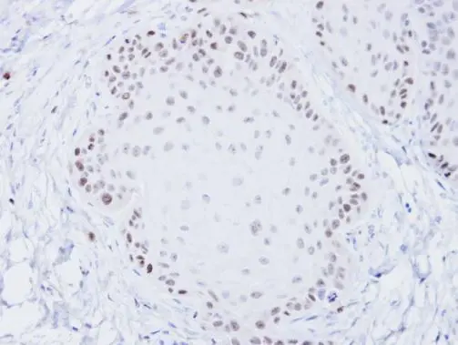
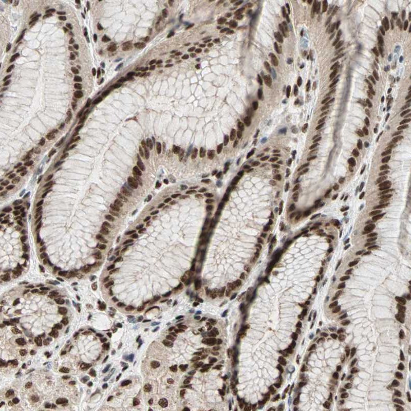
Immunohistochemical analysis of paraffin-embedded Cal27 xenograft, using BLM(GTX101303) antibody at 1:100 dilution.
Antigen Retrieval: Trilogy? (EDTA based, pH 8.0) buffer, 15min
![Untreated (–) and treated (+) THP-1 cells were untreated or treated with 10 ng/ml PMA for 24 hrs (30 μg) were separated by 5% SDS-PAGE, and the membrane was blotted with BLM antibody [C3], C-term (GTX101303) diluted at 1:1000. The HRP-conjugated anti-rabbit IgG antibody (GTX213110-01) was used to detect the primary antibody, and the signal was developed with Trident ECL plus-Enhanced.](https://www.genetex.com/upload/website/prouct_img/normal/GTX101303/GTX101303_44489_20211217_WB_treatment_PMA_w_23060100_595.webp "Untreated (–) and treated (+) THP-1 cells were untreated or treated with 10 ng/ml PMA for 24 hrs (30 μg) were separated by 5% SDS-PAGE, and the membrane was blotted with BLM antibody [C3], C-term (GTX101303) diluted at 1:1000. The HRP-conjugated anti-rabbit IgG antibody (GTX213110-01) was used to detect the primary antibody, and the signal was developed with Trident ECL plus-Enhanced.")
![BLM antibody [C3], C-term detects BLM protein at nucleus by immunofluorescent analysis. Sample: A431 cells were fixed in 4% paraformaldehyde at RT for 15 min. Green: BLM protein stained by BLM antibody [C3], C-term (GTX101303) diluted at 1:200. Blue: Hoechst 33342 staining.](https://www.genetex.com/upload/website/prouct_img/normal/GTX101303/GTX101303_39862_20160525_IFA_w_23060100_140.webp "BLM antibody [C3], C-term detects BLM protein at nucleus by immunofluorescent analysis. Sample: A431 cells were fixed in 4% paraformaldehyde at RT for 15 min. Green: BLM protein stained by BLM antibody [C3], C-term (GTX101303) diluted at 1:200. Blue: Hoechst 33342 staining.")
![Various whole cell extracts (30 μg) were separated by 5% SDS-PAGE, and the membrane was blotted with BLM antibody [C3], C-term (GTX101303) diluted at 1:1000. The HRP-conjugated anti-rabbit IgG antibody (GTX213110-01) was used to detect the primary antibody. Corresponding RNA expression data for the same cell lines are based on Human Protein Atlas program.](https://www.genetex.com/upload/website/prouct_img/normal/GTX101303/GTX101303_44489_20240202_WB_TPM_watermark_24021917_826.webp "Various whole cell extracts (30 μg) were separated by 5% SDS-PAGE, and the membrane was blotted with BLM antibody [C3], C-term (GTX101303) diluted at 1:1000. The HRP-conjugated anti-rabbit IgG antibody (GTX213110-01) was used to detect the primary antibody. Corresponding RNA expression data for the same cell lines are based on Human Protein Atlas program.")
Immunohistochemical analysis of paraffin-embedded Cal27 xenograft, using BLM(GTX101303) antibody at 1:100 dilution.
Antigen Retrieval: Trilogy? (EDTA based, pH 8.0) buffer, 15min
BLM antibody [C3], C-term
GTX101303
ApplicationsImmunoFluorescence, Western Blot, ImmunoCytoChemistry, ImmunoHistoChemistry, ImmunoHistoChemistry Paraffin
Product group Antibodies
ReactivityHuman
TargetBLM
Overview
- SupplierGeneTex
- Product NameBLM antibody [C3], C-term
- Delivery Days Customer9
- Application Supplier NoteWB: 1:500-1:3000. ICC/IF: 1:100-1:1000. IHC-P: 1:100-1:1000. *Optimal dilutions/concentrations should be determined by the researcher.Not tested in other applications.
- ApplicationsImmunoFluorescence, Western Blot, ImmunoCytoChemistry, ImmunoHistoChemistry, ImmunoHistoChemistry Paraffin
- CertificationResearch Use Only
- ClonalityPolyclonal
- Concentration1.34 mg/ml
- ConjugateUnconjugated
- Gene ID641
- Target nameBLM
- Target descriptionBLM RecQ like helicase
- Target synonymsBS, MGRISCE1, RECQ2, RECQL2, RECQL3, recQ-like DNA helicase BLM, Bloom syndrome RecQ like helicase, Bloom syndrome, RecQ helicase-like, DNA 3'-5' helicase BLM, DNA helicase, RecQ-like type 2, bloom syndrome protein, recQ protein-like 3
- HostRabbit
- IsotypeIgG
- Protein IDP54132
- Protein NameRecQ-like DNA helicase BLM
- Scientific DescriptionThe Bloom syndrome gene product is related to the RecQ subset of DExH box-containing DNA helicases and has both DNA-stimulated ATPase and ATP-dependent DNA helicase activities. Mutations causing Bloom syndrome delete or alter helicase motifs and may disable the 3-5 helicase activity. The normal protein may act to suppress inappropriate recombination. [provided by RefSeq]
- ReactivityHuman
- Storage Instruction-20°C or -80°C,2°C to 8°C
- UNSPSC41116161
Datasheet
Related products
Product group Antibodies
ApplicationsImmunoFluorescence, ELISA, ImmunoHistoChemistry
ReactivityHuman, Mouse
TargetBLM
- SizePrice
Product group Antibodies

ApplicationsImmunoFluorescence, Western Blot, ELISA, ImmunoCytoChemistry, ImmunoHistoChemistry
- SizePrice
Product group Antibodies
Anti-BLM AntibodyA47280
ApplicationsImmunoHistoChemistry
ReactivityHuman
- SizePrice
Product group Antibodies
Anti-BLM AntibodyHPA005689
ApplicationsImmunoCytoChemistry, ImmunoHistoChemistry
ReactivityHuman
TargetBLM
- SizePrice
Product group Antibodies
Phospho-BLM (T99) AntibodyCSB-PA050012
ApplicationsImmunoFluorescence, Western Blot, ELISA, ImmunoHistoChemistry
ReactivityHuman
TargetBLM
- SizePrice
Product group Antibodies

BLM Antibody (aa1375-C-term)LS-C287043
ApplicationsImmunoPrecipitation, Western Blot
ReactivityHuman, Mouse
TargetBLM
- SizePrice
Product group Antibodies
BLM antibodyGTX25409
ApplicationsWestern Blot, ImmunoHistoChemistry, ImmunoHistoChemistry Frozen
ReactivityHuman
TargetBLM
- SizePrice
![Untreated (–) and treated (+) THP-1 whole cell extracts (30 μg) were separated by 5% SDS-PAGE, and the membrane was blotted with BLM antibody [HL2771] (GTX639636) diluted at 1:1000. The HRP-conjugated anti-rabbit IgG antibody (GTX213110-01) was used to detect the primary antibody.](https://www.genetex.com/upload/website/prouct_img/normal/GTX639636/GTX639636_T-45299_20240126_WB_treatment_PMA_24013018_522.webp)
Product group Antibodies
BLM antibody [HL2771]GTX639636
ApplicationsWestern Blot
ReactivityHuman
TargetBLM
- SizePrice
![BLM antibody [N1N2], N-term detects BLM protein at nucleus by immunofluorescent analysis. Sample: HeLa cells were fixed in 4% paraformaldehyde at RT for 15 min. Green: BLM stained by BLM antibody [N1N2], N-term (GTX101403) diluted at 1:500. Red: alpha Tubulin, a cytoskeleton marker, stained by alpha Tubulin antibody [GT114] (GTX628802) diluted at 1:1000.](https://www.genetex.com/upload/website/prouct_img/normal/GTX101403/GTX101403_41584_20220218_ICC_IF_w_23060100_842.webp)
Product group Antibodies
BLM antibody [N1N2], N-termGTX101403
ApplicationsImmunoFluorescence, Western Blot, ImmunoCytoChemistry
ReactivityHuman
TargetBLM
- SizePrice
![Whole cell extract (30 μg) was separated by 5% SDS-PAGE, and the membrane was blotted with BLM antibody [N1N3] (GTX108868) diluted at 1:1000. The HRP-conjugated anti-rabbit IgG antibody (GTX213110-01) was used to detect the primary antibody.](https://www.genetex.com/upload/website/prouct_img/normal/GTX108868/GTX108868_40450_20220311_WB_w_23060120_512.webp)
Product group Antibodies
BLM antibody [N1N3]GTX108868
ApplicationsWestern Blot
ReactivityHuman
TargetBLM
- SizePrice