

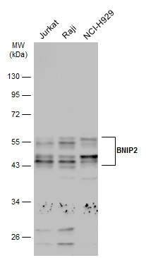
Various whole cell extracts (30 μg) were separated by 10% SDS-PAGE, and the membrane was blotted with BNIP2 antibody (GTX114283) diluted at 1:1000. The HRP-conjugated anti-rabbit IgG antibody (GTX213110-01) was used to detect the primary antibody.
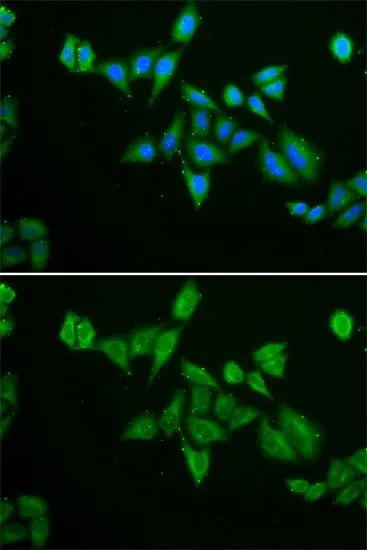
![BNIP2 antibody detects BNIP2 protein by immunofluorescent analysis. Sample: DIV10 rat E18 primary cortical neurons were fixed in 4% paraformaldehyde at RT for 15 min. Green: BNIP2 protein stained by BNIP2 antibody (GTX114283) diluted at 1:500. Red: beta Tubulin 3/ Tuj1, stained by beta Tubulin 3/ Tuj1 antibody [GT1338] (GTX631831) diluted at 1:500. Blue: Fluoroshield with DAPI (GTX30920).](https://www.genetex.com/upload/website/prouct_img/normal/GTX114283/GTX114283_40702_20170824_IFA_w_23060501_243.webp "BNIP2 antibody detects BNIP2 protein by immunofluorescent analysis. Sample: DIV10 rat E18 primary cortical neurons were fixed in 4% paraformaldehyde at RT for 15 min. Green: BNIP2 protein stained by BNIP2 antibody (GTX114283) diluted at 1:500. Red: beta Tubulin 3/ Tuj1, stained by beta Tubulin 3/ Tuj1 antibody [GT1338] (GTX631831) diluted at 1:500. Blue: Fluoroshield with DAPI (GTX30920).")
 antibody at 1:500 dilution.
Antigen Retrieval: Trilogy? (EDTA based, pH 8.0) buffer, 15min")
Various whole cell extracts (30 μg) were separated by 10% SDS-PAGE, and the membrane was blotted with BNIP2 antibody (GTX114283) diluted at 1:1000. The HRP-conjugated anti-rabbit IgG antibody (GTX213110-01) was used to detect the primary antibody.
BNIP2 antibody
GTX114283
ApplicationsImmunoFluorescence, Western Blot, ImmunoCytoChemistry, ImmunoHistoChemistry, ImmunoHistoChemistry Paraffin
Product group Antibodies
ReactivityHuman, Rat
TargetBNIP2
Overview
- SupplierGeneTex
- Product NameBNIP2 antibody
- Delivery Days Customer9
- Application Supplier NoteWB: 1:500-1:3000. ICC/IF: 1:100-1:1000. IHC-P: 1:100-1:1000. *Optimal dilutions/concentrations should be determined by the researcher.Not tested in other applications.
- ApplicationsImmunoFluorescence, Western Blot, ImmunoCytoChemistry, ImmunoHistoChemistry, ImmunoHistoChemistry Paraffin
- CertificationResearch Use Only
- ClonalityPolyclonal
- Concentration0.95 mg/ml
- ConjugateUnconjugated
- Gene ID663
- Target nameBNIP2
- Target descriptionBCL2 interacting protein 2
- Target synonymsBNIP-2, NIP2, BCL2/adenovirus E1B 19 kDa protein-interacting protein 2, BCL2/adenovirus E1B 19kDa interacting protein 2
- HostRabbit
- IsotypeIgG
- Protein IDQ12982
- Protein NameBCL2/adenovirus E1B 19 kDa protein-interacting protein 2
- Scientific DescriptionThis gene is a member of the BCL2/adenovirus E1B 19 kd-interacting protein (BNIP) family. Though the specific function is unknown, it interacts with the E1B 19 kDa protein which is responsible for the protection of virally-induced cell death, as well as E1B 19 kDa-like sequences of BCL2, also an apoptotic protector. [provided by RefSeq]
- ReactivityHuman, Rat
- Storage Instruction-20°C or -80°C,2°C to 8°C
- UNSPSC41116161
Datasheet
Related products
Product group Antibodies
BNIP2 AntibodyCSB-PA001039
ApplicationsWestern Blot, ELISA, ImmunoHistoChemistry
ReactivityHuman, Mouse
TargetBNIP2
- SizePrice
Product group Antibodies
Anti-BNIP2 AntibodyA101195
ApplicationsWestern Blot, ELISA
ReactivityHuman
- SizePrice

Product group Antibodies
Anti-BNIP-2 AntibodyA07336
ApplicationsImmunoFluorescence, Western Blot, ELISA, ImmunoHistoChemistry
ReactivityHuman, Mouse
TargetBNIP2
- SizePrice

Product group Antibodies
Anti-BNIP2 AntibodyHPA026843
ApplicationsWestern Blot, ImmunoHistoChemistry
ReactivityHuman, Rat
TargetBNIP2
- SizePrice
Product group Antibodies

BNIP2 AntibodyLS-C406222
ApplicationsELISA, ImmunoHistoChemistry
ReactivityHuman, Mouse
TargetBNIP2
- SizePrice
Product group Antibodies
References
BNIP2 antibodyGTX30091
ApplicationsImmunoFluorescence, Western Blot, ImmunoCytoChemistry
ReactivityHuman, Mouse
TargetBNIP2
- SizePrice

Product group Antibodies
BNIP2 antibodyGTX87240
ApplicationsWestern Blot
ReactivityHuman
TargetBNIP2
- SizePrice
Product group Antibodies
Anti-BNIP2 Antibody144-06282
ApplicationsImmunoFluorescence, Western Blot
ReactivityHuman, Mouse
TargetBNIP2
- SizePrice