

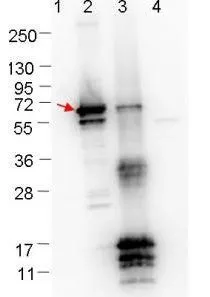
Western blot showing detection of 0.1 μg recombinant proteins in Western blot. Lane 1: Molecular weight markers. Lane 2: MBP-ErpN/OspE fusion protein (arrow; 59.5 kDa expected MW). Lane 3: fusion protein (MBP-tagged) plus cleaved fusion proteins (without MBP). Lane 4: MBP alone. The lower bands are probably breakdown products. The upper bands in lane 3 are fusion protein (top band), or breakdown products of the fusion protein (bands in middle of blot). Protein was run on a 4-20% gel, then transferred to 0.45 μm nitrocellulose. After blocking with 1% BSA-TTBS overnight at 4oC, primary antibody was used at 1:1000 at room temperature for 30 min. HRP-conjugated Goat-Anti-Rabbit secondary antibody was used at 1:40,000 in blocking buffer and imaged on the VersaDoc MP 4000 imaging system (Bio-Rad).
Western blot showing detection of 0.1 μg recombinant proteins in Western blot. Lane 1: Molecular weight markers. Lane 2: MBP-ErpN/OspE fusion protein (arrow; 59.5 kDa expected MW). Lane 3: fusion protein (MBP-tagged) plus cleaved fusion proteins (without MBP). Lane 4: MBP alone. The lower bands are probably breakdown products. The upper bands in lane 3 are fusion protein (top band), or breakdown products of the fusion protein (bands in middle of blot). Protein was run on a 4-20% gel, then transferred to 0.45 μm nitrocellulose. After blocking with 1% BSA-TTBS overnight at 4oC, primary antibody was used at 1:1000 at room temperature for 30 min. HRP-conjugated Goat-Anti-Rabbit secondary antibody was used at 1:40,000 in blocking buffer and imaged on the VersaDoc MP 4000 imaging system (Bio-Rad).
Borrelia burgdorferi Erpn/Ospe antibody
GTX48807
ApplicationsWestern Blot, ELISA
Product group Antibodies
ReactivityBacteria
TargeterpA
Overview
- SupplierGeneTex
- Product NameBorrelia burgdorferi Erpn/Ospe antibody
- Delivery Days Customer9
- Application Supplier NoteWB: 1:1000. ELISA: 1:5000. *Optimal dilutions/concentrations should be determined by the researcher.Not tested in other applications.
- ApplicationsWestern Blot, ELISA
- CertificationResearch Use Only
- ClonalityPolyclonal
- Concentration1 mg/ml
- ConjugateUnconjugated
- Gene ID1194664
- Target nameerpA
- Target descriptionplasminogen-binding protein ErpA
- Target synonymsBB_RS07085, BB_L39, plasminogen-binding protein ErpA
- HostRabbit
- IsotypeIgG
- ReactivityBacteria
- Storage Instruction-20°C or -80°C,2°C to 8°C
- UNSPSC41116161