

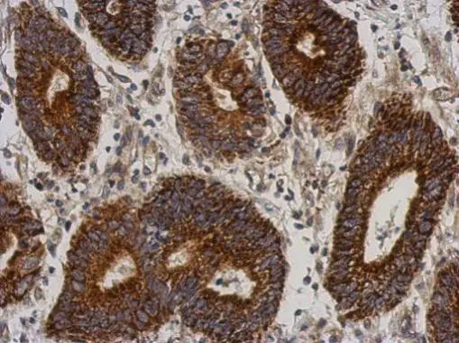
Immunohistochemical analysis of paraffin-embedded human colon carcinoma, using C21orf2(GTX119046) antibody at 1:500 dilution.
Antigen Retrieval: Trilogy? (EDTA based, pH 8.0) buffer, 15min
![C21orf2 antibody [N1C2] detects C21orf2 protein at cytoplasm and nucleus by immunofluorescent analysis. Sample: A431 cells were fixed in 4% paraformaldehyde at RT for 15 min. Green: C21orf2 stained by C21orf2 antibody [N1C2] (GTX119046) diluted at 1:500. Blue: Hoechst 33342 staining. Scale bar= 10 μm.](https://www.genetex.com/upload/website/prouct_img/normal/GTX119046/GTX119046_40310_20190306_ICC_IF_w_23060519_346.webp "C21orf2 antibody [N1C2] detects C21orf2 protein at cytoplasm and nucleus by immunofluorescent analysis. Sample: A431 cells were fixed in 4% paraformaldehyde at RT for 15 min. Green: C21orf2 stained by C21orf2 antibody [N1C2] (GTX119046) diluted at 1:500. Blue: Hoechst 33342 staining. Scale bar= 10 μm.")
 A: Jurkat 10% SDS PAGE GTX119046 diluted at 1:500")
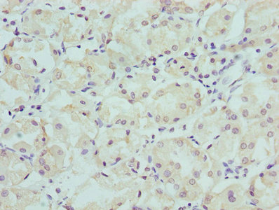
Immunohistochemical analysis of paraffin-embedded human colon carcinoma, using C21orf2(GTX119046) antibody at 1:500 dilution.
Antigen Retrieval: Trilogy? (EDTA based, pH 8.0) buffer, 15min
C21orf2 antibody [N1C2]
GTX119046
ApplicationsImmunoFluorescence, Western Blot, ImmunoCytoChemistry, ImmunoHistoChemistry, ImmunoHistoChemistry Paraffin
Product group Antibodies
ReactivityHuman
TargetCFAP410
Overview
- SupplierGeneTex
- Product NameC21orf2 antibody [N1C2]
- Delivery Days Customer9
- Application Supplier NoteWB: 1:500-1:3000. ICC/IF: 1:100-1:1000. IHC-P: 1:100-1:1000. *Optimal dilutions/concentrations should be determined by the researcher.Not tested in other applications.
- ApplicationsImmunoFluorescence, Western Blot, ImmunoCytoChemistry, ImmunoHistoChemistry, ImmunoHistoChemistry Paraffin
- CertificationResearch Use Only
- ClonalityPolyclonal
- Concentration0.72 mg/ml
- ConjugateUnconjugated
- Gene ID755
- Target nameCFAP410
- Target descriptioncilia and flagella associated protein 410
- Target synonymsC21orf2, LRRC76, RDMS, SMDAX, YF5/A2, cilia- and flagella-associated protein 410, nuclear encoded mitochondrial protein C21orf2, C21orf-HUMF09G8.5, leucine rich repeat containing 76, leucine-rich repeat-containing protein 76, protein C21orf2
- HostRabbit
- IsotypeIgG
- Protein IDO43822
- Protein NameCilia- and flagella-associated protein 410
- ReactivityHuman
- Storage Instruction-20°C or -80°C,2°C to 8°C
- UNSPSC41116161
Datasheet
Related products
Product group Antibodies
Anti-CU002 (C-term) Antibody102-20619
ApplicationsFlow Cytometry, Western Blot
TargetCFAP410
- SizePrice

Product group Antibodies
CFAP410 AntibodyCSB-PA003758LA01HU
ApplicationsELISA, ImmunoHistoChemistry
ReactivityHuman
TargetCFAP410
- SizePrice
Product group Antibodies
Cfap410 Polyclonal AntibodyCAC11773
ApplicationsELISA, ImmunoHistoChemistry
TargetCFAP410
- SizePrice

Product group Antibodies
Anti-C21orf2 AntibodyHPA030284
ApplicationsImmunoHistoChemistry
ReactivityHuman
TargetCFAP410
- SizePrice
Product group Antibodies

C21orf2 AntibodyLS-C396219
ApplicationsELISA
ReactivityHuman
TargetCFAP410
- SizePrice
Product group Antibodies
CFAP410 AntibodyPACO25352
ApplicationsELISA, ImmunoHistoChemistry
ReactivityHuman
TargetCFAP410
- SizePrice