

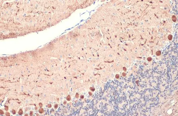
Calbindin antibody detects Calbindin protein at cytoplasm by immunohistochemical analysis. Sample: Paraffin-embedded mouse cerebellum. Calbindin stained by Calbindin antibody (GTX130856) diluted at 1:1000. Antigen Retrieval: Citrate buffer, pH 6.0, 15 min
 diluted at 1:1000. Antigen Retrieval: Citrate buffer, pH 6.0, 15 min")
 were separated by 12% SDS-PAGE, and the membrane was blotted with Calbindin antibody (GTX130856) diluted at 1:1000. The HRP-conjugated anti-rabbit IgG antibody (GTX213110-01) was used to detect the primary antibody.")
 was separated by 12% SDS-PAGE, and the membrane was blotted with Calbindin antibody (GTX130856) diluted at 1:1000. The HRP-conjugated anti-rabbit IgG antibody (GTX213110-01) was used to detect the primary antibody.")
![Calbindin antibody detects Calbindin protein by immunofluorescent analysis. Sample: DIV9 rat E18 primary cortical neuron cells were fixed in 4% paraformaldehyde at RT for 15 min. Green: Calbindin stained by Calbindin antibody (GTX130856) diluted at 1:500. Red: beta Tubulin 3/ Tuj1, stained by beta Tubulin 3/ Tuj1 antibody [GT1338] (GTX631831) diluted at 1:500. Blue: Fluoroshield with DAPI (GTX30920).](https://www.genetex.com/upload/website/prouct_img/normal/GTX130856/GTX130856_42158_20171115_ICC_IF_R_w_23060523_862.webp "Calbindin antibody detects Calbindin protein by immunofluorescent analysis. Sample: DIV9 rat E18 primary cortical neuron cells were fixed in 4% paraformaldehyde at RT for 15 min. Green: Calbindin stained by Calbindin antibody (GTX130856) diluted at 1:500. Red: beta Tubulin 3/ Tuj1, stained by beta Tubulin 3/ Tuj1 antibody [GT1338] (GTX631831) diluted at 1:500. Blue: Fluoroshield with DAPI (GTX30920).")
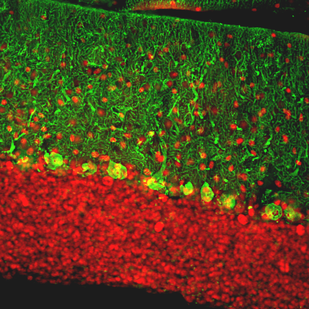
![Calbindin antibody detects Calbindin protein expression by immunohistochemical analysis. Sample: Frozen-sectioned adult mouse cerebellum. Green: Calbindin protein stained by Calbindin antibody (GTX130856) diluted at 1:250. Red: beta Tubulin 3/ TUJ1, stained by beta Tubulin 3/ TUJ1 antibody [GT11710] (GTX631836) diluted at 1:500. Blue: Fluoroshield with DAPI (GTX30920).](https://www.genetex.com/upload/website/prouct_img/normal/GTX130856/GTX130856_42158_20170531_IHC-Fr_M_1_w_23060523_743.webp "Calbindin antibody detects Calbindin protein expression by immunohistochemical analysis. Sample: Frozen-sectioned adult mouse cerebellum. Green: Calbindin protein stained by Calbindin antibody (GTX130856) diluted at 1:250. Red: beta Tubulin 3/ TUJ1, stained by beta Tubulin 3/ TUJ1 antibody [GT11710] (GTX631836) diluted at 1:500. Blue: Fluoroshield with DAPI (GTX30920).")
 were separated by 12% SDS-PAGE, and the membranes were blotted with Calbindin antibody (GTX130856) diluted at 1:1000 and competitor's antibody (sc-7691) diluted at 1:1000. The HRP-conjugated anti-rabbit IgG antibody (GTX213110-01) was used to detect the primary antibody.")
![Calbindin antibody detects Calbindin protein expression by immunohistochemical analysis. Sample: Frozen-sectioned adult mouse cerebellum. Green: Calbindin protein stained by Calbindin antibody (GTX130856) diluted at 1:250. Red: beta Tubulin 3/ TUJ1, stained by beta Tubulin 3/ TUJ1 antibody [GT11710] (GTX631836) diluted at 1:500. Blue: Fluoroshield with DAPI (GTX30920).](https://www.genetex.com/upload/website/prouct_img/normal/GTX130856/GTX130856_42158_20170531_IHC-Fr_M_2_w_23060523_647.webp "Calbindin antibody detects Calbindin protein expression by immunohistochemical analysis. Sample: Frozen-sectioned adult mouse cerebellum. Green: Calbindin protein stained by Calbindin antibody (GTX130856) diluted at 1:250. Red: beta Tubulin 3/ TUJ1, stained by beta Tubulin 3/ TUJ1 antibody [GT11710] (GTX631836) diluted at 1:500. Blue: Fluoroshield with DAPI (GTX30920).")
 diluted at 1:1000.
Antigen Retrieval: Citrate buffer, pH 6.0, 15 min")
![Calbindin antibody detects Calbindin protein by immunohistochemical analysis. Samples: Paraffin-embedded mouse retina. Green: Calbindin protein stained by Calbindin antibody (GTX130856) diluted at 1:500. Red: beta Tubulin 3/ Tuj1, a marker, stained by beta Tubulin 3/ Tuj1 antibody [GT886] (GTX631830) diluted at 1:500. Blue: Fluoroshield with DAPI (GTX30920).
Antigen Retrieval: Citrate buffer, pH 6.0, 15 min](https://www.genetex.com/upload/website/prouct_img/normal/GTX130856/GTX130856_42158_20171127_IHC-P_M_w_23060523_987.webp "Calbindin antibody detects Calbindin protein by immunohistochemical analysis. Samples: Paraffin-embedded mouse retina. Green: Calbindin protein stained by Calbindin antibody (GTX130856) diluted at 1:500. Red: beta Tubulin 3/ Tuj1, a marker, stained by beta Tubulin 3/ Tuj1 antibody [GT886] (GTX631830) diluted at 1:500. Blue: Fluoroshield with DAPI (GTX30920).
Antigen Retrieval: Citrate buffer, pH 6.0, 15 min")
Calbindin antibody detects Calbindin protein at cytoplasm by immunohistochemical analysis. Sample: Paraffin-embedded mouse cerebellum. Calbindin stained by Calbindin antibody (GTX130856) diluted at 1:1000. Antigen Retrieval: Citrate buffer, pH 6.0, 15 min
Calbindin antibody
GTX130856
ApplicationsImmunoFluorescence, Western Blot, ImmunoCytoChemistry, ImmunoHistoChemistry, ImmunoHistoChemistry Frozen, ImmunoHistoChemistry Paraffin
Product group Antibodies
ReactivityHuman, Mouse, Rat
TargetCALB1
Overview
- SupplierGeneTex
- Product NameCalbindin antibody
- Delivery Days Customer9
- Application Supplier NoteWB: 1:500-1:3000. ICC/IF: 1:100-1:1000. IHC-P: 1:100-1:1000. IHC-Fr: 1:100-1:1000. *Optimal dilutions/concentrations should be determined by the researcher.Not tested in other applications.
- ApplicationsImmunoFluorescence, Western Blot, ImmunoCytoChemistry, ImmunoHistoChemistry, ImmunoHistoChemistry Frozen, ImmunoHistoChemistry Paraffin
- CertificationResearch Use Only
- ClonalityPolyclonal
- Concentration0.51 mg/ml
- ConjugateUnconjugated
- Gene ID793
- Target nameCALB1
- Target descriptioncalbindin 1
- Target synonymsCALB, D-28K, calbindin, RTVL-H protein, calbindin 1, (28kD), calbindin 1, 28kDa, calbindin 27, calbindin D28, calbindin-D28k, vitamin D-dependent calcium-binding protein, avian-type
- HostRabbit
- IsotypeIgG
- Protein IDP05937
- Protein NameCalbindin
- Scientific DescriptionCalbindin is a calcium-binding protein belonging to the troponin C superfamily. It was originally described as a 27-kD protein induced by vitamin D in the duodenum of the chick. In the brain, its synthesis is independent of vitamin-D-derived hormones. Calbindin contains 4 active calcium-binding domains, and 2 modified domains that presumably have lost their calcium-binding capacity. The neurons in brains of patients with Huntington disease are calbindin-depleted. [provided by RefSeq]
- ReactivityHuman, Mouse, Rat
- Storage Instruction-20°C or -80°C,2°C to 8°C
- UNSPSC41116161
Datasheet
Related products
Product group Antibodies
Anti-Calbindin AntibodyA85359
ApplicationsImmunoFluorescence, Western Blot, ImmunoCytoChemistry, ImmunoHistoChemistry
ReactivityBovine, Human, Mouse, Rat
- SizePrice
Product group Antibodies
Anti-Calbindin/CALB1 Antibody Picoband(r)A03047-CARRIER-FREE
ApplicationsFlow Cytometry, ImmunoFluorescence, Western Blot, ELISA, ImmunoHistoChemistry
ReactivityHuman, Mouse, Rat
TargetCALB1
- SizePrice
Product group Antibodies
Anti-CALB1 Antibody144-60033
ApplicationsWestern Blot, ImmunoHistoChemistry
ReactivityHuman, Mouse, Rat
TargetCALB1
- SizePrice
Product group Antibodies

Calbindin 1 (CALB1) AntibodyABX034864
ApplicationsWestern Blot, ELISA, ImmunoHistoChemistry
- SizePrice
Product group Antibodies

CALB1 / Calbindin AntibodyLS-C829963
ApplicationsELISA, ImmunoHistoChemistry
ReactivityHuman, Mouse, Rat
TargetCALB1
- SizePrice
Product group Antibodies
CALB1 Recombinant AntibodyBSM-60713R
ApplicationsImmunoFluorescence, Western Blot, ImmunoHistoChemistry, ImmunoHistoChemistry Frozen, ImmunoHistoChemistry Paraffin
ReactivityHuman, Mouse, Rat
TargetCALB1
- SizePrice
Product group Antibodies
CALB1 AntibodyCSB-PA004432LA01HU
ApplicationsImmunoFluorescence, ELISA, ImmunoHistoChemistry
ReactivityHuman
TargetCALB1
- SizePrice
Product group Antibodies

Goat anti-calbindin D28EB11759
ApplicationsWestern Blot, ELISA, ImmunoHistoChemistry
ReactivityBovine, Canine, Human, Mouse, Porcine, Rat
TargetCALB1
- SizePrice
Product group Antibodies
ApplicationsImmunoPrecipitation, Western Blot, ImmunoCytoChemistry, ImmunoHistoChemistry
ReactivityMouse, Porcine
TargetCALB1
- SizePrice
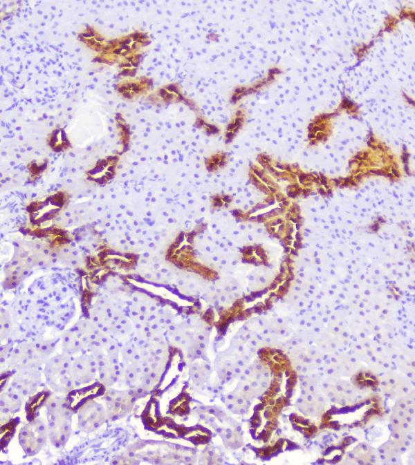
![IHC-P analysis of human renal cortex tissue using GTX04454 Calbindin antibody [MSVA-471M] HistoMAX?. A strong cytoplasmic calbindin immunostaining occurs in a fraction of distal tubuli of the kidney.](https://www.genetex.com/upload/website/prouct_img/normal/GTX04454/GTX04454_20230728_IHC-P_7_23072722_747.webp)
Product group Antibodies
ApplicationsImmunoHistoChemistry, ImmunoHistoChemistry Paraffin
ReactivityHuman
TargetCALB1
- SizePrice